 Movie
Movie Controller

[English] 日本語
 Yorodumi
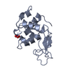
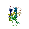
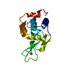
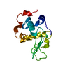
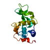
- PDB-1ljf: CRYSTAL STRUCTURE OF MONOCLINIC LYSOZYME GROWN IN PRESENCE OF 10%... -
+ Open data

- Basic information
| Entry | Database: PDB / ID: 1ljf | ||||||
|---|---|---|---|---|---|---|---|
| Title | CRYSTAL STRUCTURE OF MONOCLINIC LYSOZYME GROWN IN PRESENCE OF 10% SUCROSE | ||||||
 Components Components | Lysozyme C | ||||||
 Keywords Keywords |  HYDROLASE / Hydration of Proteins HYDROLASE / Hydration of Proteins | ||||||
| Function / homology |  Function and homology informationAntimicrobial peptides / Neutrophil degranulation / beta-N-acetylglucosaminidase activity / cell wall macromolecule catabolic process / lysozyme / lysozyme activity / killing of cells of another organism / defense response to Gram-negative bacterium / defense response to Gram-positive bacterium / defense response to bacterium ...Antimicrobial peptides / Neutrophil degranulation / beta-N-acetylglucosaminidase activity / cell wall macromolecule catabolic process / lysozyme / lysozyme activity / killing of cells of another organism / defense response to Gram-negative bacterium / defense response to Gram-positive bacterium / defense response to bacterium / endoplasmic reticulum / extracellular space / identical protein binding / cytoplasm Function and homology informationAntimicrobial peptides / Neutrophil degranulation / beta-N-acetylglucosaminidase activity / cell wall macromolecule catabolic process / lysozyme / lysozyme activity / killing of cells of another organism / defense response to Gram-negative bacterium / defense response to Gram-positive bacterium / defense response to bacterium ...Antimicrobial peptides / Neutrophil degranulation / beta-N-acetylglucosaminidase activity / cell wall macromolecule catabolic process / lysozyme / lysozyme activity / killing of cells of another organism / defense response to Gram-negative bacterium / defense response to Gram-positive bacterium / defense response to bacterium / endoplasmic reticulum / extracellular space / identical protein binding / cytoplasmSimilarity search - Function | ||||||
| Biological species |  Gallus gallus (chicken) Gallus gallus (chicken) | ||||||
| Method | X-RAY DIFFRACTION / Resolution: 1.8 Å | ||||||
 Authors Authors | Saraswathi, N.T. / Sankaranarayanan, R. / Vijayan, M. | ||||||
 Citation Citation | Journal: Acta Crystallogr.,Sect.D / Year: 2002 Title: Effect of stabilizing additives on the structure and hydration of proteins: a study involving monoclinic lysozyme. Authors: Saraswathi, N.T. / Sankaranarayanan, R. / Vijayan, M. #1: Journal: Acta Crystallogr.,Sect.D / Year: 2001Title: The Effect of Stabilizing Additives on the Structure and Hydration of Proteins: A Study Involving Tetragonal Lysozyme Authors: Datta, S. / Biswal, B.K. / Vijayan, M. #2: Journal: Acta Crystallogr.,Sect.D / Year: 1996Title: An X-Ray Analysis of Native Monoclinic Lysozyme. A Case Study on the Reliability of Refined Protein Structures and a Comparison with the Low-Humidity Form in Relation to Mobility and Enzyme Action Authors: Nagendra, H.G. / Sudarsanakumar, C. / Vijayan, M. | ||||||
| History |
|
- Structure visualization
| Structure viewer | Molecule: MolmilJmol/JSmol |
|---|
- Downloads & links
-Download
| PDBx/mmCIF format | 1ljf.cif.gz | 65.5 KB | Display | PDBx/mmCIF format |
|---|---|---|---|---|
| PDB format | pdb1ljf.ent.gz | 52.2 KB | Display | PDB format |
| PDBx/mmJSON format | 1ljf.json.gz | Tree view | PDBx/mmJSON format | |
| Others |  Other downloads Other downloads |
-Validation report
| Arichive directory | https://data.pdbj.org/pub/pdb/validation_reports/lj/1ljfftp://data.pdbj.org/pub/pdb/validation_reports/lj/1ljf | HTTPS FTP |
|---|
-Related structure data
| Related structure data | 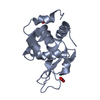 1lj3C 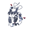 1lj4C 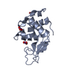 1ljeC 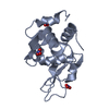 1ljgC 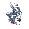 1ljhC 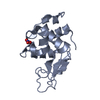 1ljiC 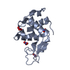 1ljjC 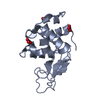 1ljkC C: citing same article ( |
|---|---|
| Similar structure data |


-Links
 PDBj
PDBj


- Assembly
| Deposited unit | 
| ||||||||||
|---|---|---|---|---|---|---|---|---|---|---|---|
| 1 | 
| ||||||||||
| 2 | 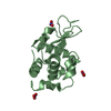
| ||||||||||
| Unit cell |
|
-Components
| #1: Protein | Mass: 14331.160 Da / Num. of mol.: 2 / Source method: isolated from a natural source / Source: (natural) Gallus gallus (chicken) / References: UniProt: P00698, lysozyme#2: Chemical | ChemComp-NO3 / Nitrate  Mass: 62.005 Da / Num. of mol.: 6 / Source method: obtained synthetically / Formula: NO3 Mass: 62.005 Da / Num. of mol.: 6 / Source method: obtained synthetically / Formula: NO3#3: Water | ChemComp-HOH / | Water Mass: 18.015 Da / Num. of mol.: 349 / Source method: isolated from a natural source / Formula: H2O Mass: 18.015 Da / Num. of mol.: 349 / Source method: isolated from a natural source / Formula: H2O |
|---|
-Experimental details
-Experiment
| Experiment | Method: X-RAY DIFFRACTION / Number of used crystals: 1 |
|---|
- Sample preparation
| Crystal | Density Matthews: 1.86 Å3/Da / Density % sol: 33.71 % | ||||||||||||||||||
|---|---|---|---|---|---|---|---|---|---|---|---|---|---|---|---|---|---|---|---|
| Crystal grow | Temperature: 293 K / Method: liquid diffusion / pH: 4.6 Details: Sodium Acetate (0.1M) - Buffer, Sodium Nitrate (0.44M) - Precipitant, Sucrose (0.29M) - Additive; pH 4.6, LIQUID DIFFUSION at 293K | ||||||||||||||||||
| Crystal grow | *PLUS Method: unknown | ||||||||||||||||||
| Components of the solutions | *PLUS
|
-Data collection
| Diffraction | Mean temperature: 293 K |
|---|---|
| Diffraction source | Source: ROTATING ANODE / Type: RIGAKU RU200 / Wavelength: 1.5418 Å |
| Detector | Type: MARRESEARCH / Detector: IMAGE PLATE |
| Radiation | Protocol: SINGLE WAVELENGTH / Monochromatic (M) / Laue (L): M / Scattering type: x-ray |
| Radiation wavelength | Wavelength: 1.5418 Å / Relative weight: 1 |
| Reflection | Resolution: 1.8→30 Å / Num. obs: 18200 / Biso Wilson estimate: 21.6 Å2 |
| Reflection shell | Resolution: 1.8→1.86 Å |
| Reflection | *PLUS Highest resolution: 1.8 Å / Lowest resolution: 30 Å / Num. obs: 18248 / % possible obs: 93.4 % / Num. measured all: 135208 / Rmerge(I) obs: 0.054 |
| Reflection shell | *PLUS % possible obs: 90.9 % / Num. unique obs: 1789 / Rmerge(I) obs: 0.124 |
- Processing
| Software |
| ||||||||||||||||||||||||||||||||||||||||||||||||||||||||||||
|---|---|---|---|---|---|---|---|---|---|---|---|---|---|---|---|---|---|---|---|---|---|---|---|---|---|---|---|---|---|---|---|---|---|---|---|---|---|---|---|---|---|---|---|---|---|---|---|---|---|---|---|---|---|---|---|---|---|---|---|---|---|
| Refinement | Resolution: 1.8→30 Å / Rfactor Rfree error: 0.006 / Data cutoff high absF: 1031643.4 / Data cutoff low absF: 0 / Isotropic thermal model: RESTRAINED / Cross valid method: THROUGHOUT / σ(F): 0
| ||||||||||||||||||||||||||||||||||||||||||||||||||||||||||||
| Solvent computation | Solvent model: FLAT MODEL / Bsol: 63.2819 Å2 / ksol: 0.377077 e/Å3 | ||||||||||||||||||||||||||||||||||||||||||||||||||||||||||||
| Displacement parameters | Biso mean: 26.3 Å2
| ||||||||||||||||||||||||||||||||||||||||||||||||||||||||||||
| Refine analyze |
| ||||||||||||||||||||||||||||||||||||||||||||||||||||||||||||
| Refinement step | Cycle: LAST / Resolution: 1.8→30 Å
| ||||||||||||||||||||||||||||||||||||||||||||||||||||||||||||
| Refine LS restraints |
| ||||||||||||||||||||||||||||||||||||||||||||||||||||||||||||
| LS refinement shell | Resolution: 1.8→1.91 Å / Rfactor Rfree error: 0.02 / Total num. of bins used: 6
| ||||||||||||||||||||||||||||||||||||||||||||||||||||||||||||
| Xplor file |
| ||||||||||||||||||||||||||||||||||||||||||||||||||||||||||||
| Refinement | *PLUS Highest resolution: 1.8 Å / Lowest resolution: 30 Å | ||||||||||||||||||||||||||||||||||||||||||||||||||||||||||||
| Solvent computation | *PLUS | ||||||||||||||||||||||||||||||||||||||||||||||||||||||||||||
| Displacement parameters | *PLUS | ||||||||||||||||||||||||||||||||||||||||||||||||||||||||||||
| Refine LS restraints | *PLUS
|
