 Movie
Movie Controller

[English] 日本語
 Yorodumi
- PDB-1lfp: Crystal Structure of a Conserved Hypothetical Protein Aq1575 from... -
+ Open data

- Basic information
| Entry | Database: PDB / ID: 1lfp | ||||||
|---|---|---|---|---|---|---|---|
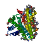
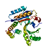
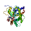
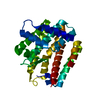
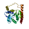
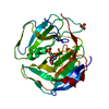
| Title | Crystal Structure of a Conserved Hypothetical Protein Aq1575 from Aquifex Aeolicus | ||||||
 Components Components | Hypothetical protein AQ_1575 Hypothesis Hypothesis | ||||||
 Keywords Keywords | RNA BINDING PROTEIN / Hypothetical / New fold / thermostability / Structural Genomics / BSGC structure funded by NIH / Protein Structure Initiative / PSI / Berkeley Structural Genomics Center | ||||||
| Function / homology |  Function and homology information Function and homology information | ||||||
| Biological species |   Aquifex aeolicus (bacteria) Aquifex aeolicus (bacteria) | ||||||
| Method | X-RAY DIFFRACTION / SYNCHROTRON / SAD / Resolution: 1.72 Å | ||||||
 Authors Authors | Shin, D.H. / Yokota, H. / Kim, R. / Kim, S.H. / Berkeley Structural Genomics Center (BSGC) | ||||||
 Citation Citation | Journal: Proc.Natl.Acad.Sci.USA / Year: 2002 Title: Crystal structure of conserved hypothetical protein Aq1575 from Aquifex aeolicus. Authors: Shin, D.H. / Yokota, H. / Kim, R. / Kim, S.H. | ||||||
| History |
|
- Structure visualization
| Structure viewer | Molecule: MolmilJmol/JSmol |
|---|
- Downloads & links
-Download
| PDBx/mmCIF format | 1lfp.cif.gz | 64.4 KB | Display | PDBx/mmCIF format |
|---|---|---|---|---|
| PDB format | pdb1lfp.ent.gz | 51.2 KB | Display | PDB format |
| PDBx/mmJSON format | 1lfp.json.gz | Tree view | PDBx/mmJSON format | |
| Others |  Other downloads Other downloads |
-Validation report
| Arichive directory | https://data.pdbj.org/pub/pdb/validation_reports/lf/1lfpftp://data.pdbj.org/pub/pdb/validation_reports/lf/1lfp | HTTPS FTP |
|---|
-Related structure data
| Similar structure data | |
|---|---|
| Other databases |


-Links
 PDBj
PDBj- Assembly
| Deposited unit | 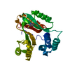
| ||||||||
|---|---|---|---|---|---|---|---|---|---|
| 1 |
| ||||||||
| Unit cell |
|
-Components
| #1: Protein | Hypothesis Mass: 28040.400 Da / Num. of mol.: 1 Source method: isolated from a genetically manipulated source Source: (gene. exp.) Aquifex aeolicus (bacteria) / Production host: Escherichia coli (E. coli) / References: UniProt: O67517 |
|---|---|
| #2: Water | ChemComp-HOH / Water Mass: 18.015 Da / Num. of mol.: 350 / Source method: isolated from a natural source / Formula: H2O Mass: 18.015 Da / Num. of mol.: 350 / Source method: isolated from a natural source / Formula: H2O |
-Experimental details
-Experiment
| Experiment | Method: X-RAY DIFFRACTION / Number of used crystals: 1 |
|---|
- Sample preparation
| Crystal | Density Matthews: 1.88 Å3/Da / Density % sol: 34.63 % | ||||||||||||||||||||||||||||||||||||||||||||||||||||||||||||||||||||||
|---|---|---|---|---|---|---|---|---|---|---|---|---|---|---|---|---|---|---|---|---|---|---|---|---|---|---|---|---|---|---|---|---|---|---|---|---|---|---|---|---|---|---|---|---|---|---|---|---|---|---|---|---|---|---|---|---|---|---|---|---|---|---|---|---|---|---|---|---|---|---|---|
| Crystal grow | Temperature: 295 K / Method: vapor diffusion, hanging drop / pH: 6.8 Details: 20% PEG3350, 0.2M Ammonium Nitrate, 10 mM NAD, 3% PEG400, pH 6.8, VAPOR DIFFUSION, HANGING DROP, temperature 295K | ||||||||||||||||||||||||||||||||||||||||||||||||||||||||||||||||||||||
| Crystal grow | *PLUS Temperature: 21.5-22.5 ℃ | ||||||||||||||||||||||||||||||||||||||||||||||||||||||||||||||||||||||
| Components of the solutions | *PLUS
|
-Data collection
| Diffraction | Mean temperature: 100 K |
|---|---|
| Diffraction source | Source: SYNCHROTRON / Site: ALS  / Beamline: 5.0.2 / Wavelength: 0.97864 Å / Beamline: 5.0.2 / Wavelength: 0.97864 Å |
| Detector | Type: ADSC QUANTUM 4 / Detector: CCD / Date: Mar 5, 2002 |
| Radiation | Monochromator: Double crystal / Protocol: SINGLE WAVELENGTH / Monochromatic (M) / Laue (L): M / Scattering type: x-ray |
| Radiation wavelength | Wavelength: 0.97864 Å / Relative weight: 1 |
| Reflection | Resolution: 1.72→36.2 Å / Num. obs: 22781 / % possible obs: 95.8 % / Observed criterion σ(F): -3 / Observed criterion σ(I): -3 / Biso Wilson estimate: 19.9 Å2 |
| Reflection shell | Resolution: 1.72→1.73 Å / % possible all: 48 |
| Reflection | *PLUS Highest resolution: 1.71 Å / Num. obs: 42899 / % possible obs: 96 % / Redundancy: 7.5 % / Rmerge(I) obs: 0.051 |
| Reflection shell | *PLUS Highest resolution: 1.71 Å / % possible obs: 48.2 % / Redundancy: 5 % / Num. unique obs: 1081 / Rmerge(I) obs: 0.207 / Mean I/σ(I) obs: 3.8 |
- Processing
| Software |
| |||||||||||||||||||||||||
|---|---|---|---|---|---|---|---|---|---|---|---|---|---|---|---|---|---|---|---|---|---|---|---|---|---|---|
| Refinement | Method to determine structure: SAD / Resolution: 1.72→29.88 Å / Rfactor Rfree error: 0.006 / Data cutoff high absF: 410192.08 / Data cutoff low absF: 0 / Isotropic thermal model: RESTRAINED / Cross valid method: THROUGHOUT / σ(F): 2 / Stereochemistry target values: Engh & Huber
| |||||||||||||||||||||||||
| Solvent computation | Solvent model: FLAT MODEL / Bsol: 45.9766 Å2 / ksol: 0.297912 e/Å3 | |||||||||||||||||||||||||
| Displacement parameters | Biso mean: 37.1 Å2
| |||||||||||||||||||||||||
| Refine analyze |
| |||||||||||||||||||||||||
| Refinement step | Cycle: LAST / Resolution: 1.72→29.88 Å
| |||||||||||||||||||||||||
| Refine LS restraints |
| |||||||||||||||||||||||||
| LS refinement shell | Resolution: 1.72→1.82 Å / Rfactor Rfree error: 0.019 / Total num. of bins used: 6
| |||||||||||||||||||||||||
| Xplor file |
| |||||||||||||||||||||||||
| Refinement | *PLUS Highest resolution: 1.71 Å / Lowest resolution: 20 Å / Rfactor obs: 0.228 | |||||||||||||||||||||||||
| Solvent computation | *PLUS | |||||||||||||||||||||||||
| Displacement parameters | *PLUS | |||||||||||||||||||||||||
| Refine LS restraints | *PLUS
| |||||||||||||||||||||||||
| LS refinement shell | *PLUS Highest resolution: 1.71 Å / Lowest resolution: 1.73 Å / Rfactor obs: 0.215 |
