 Movie
Movie Controller

[English] 日本語
 Yorodumi
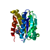
- PDB-1lf7: Crystal Structure of Human Complement Protein C8gamma at 1.2 A Re... -
+ Open data

- Basic information
| Entry | Database: PDB / ID: 1lf7 | ||||||
|---|---|---|---|---|---|---|---|
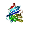
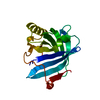
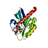
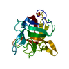
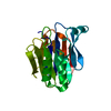
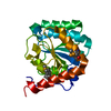
| Title | Crystal Structure of Human Complement Protein C8gamma at 1.2 A Resolution | ||||||
 Components Components | Complement Protein C8gamma | ||||||
 Keywords Keywords |  IMMUNE SYSTEM / lipocalin / beta barrel / calyx / complement / MAC IMMUNE SYSTEM / lipocalin / beta barrel / calyx / complement / MAC | ||||||
| Function / homology |  Function and homology information Function and homology informationTerminal pathway of complement / membrane attack complex / complement binding / complement activation, alternative pathway / retinol binding / complement activation, classical pathway / Regulation of Complement cascade / positive regulation of immune response / blood microparticle / killing of cells of another organism ...Terminal pathway of complement / membrane attack complex / complement binding / complement activation, alternative pathway / retinol binding / complement activation, classical pathway / Regulation of Complement cascade / positive regulation of immune response / blood microparticle / killing of cells of another organism / protein-containing complex binding / extracellular exosome / extracellular region / plasma membraneSimilarity search - Function | ||||||
| Biological species |  Homo sapiens (human) Homo sapiens (human) | ||||||
| Method | X-RAY DIFFRACTION / SYNCHROTRON / MIRAS / Resolution: 1.2 Å | ||||||
 Authors Authors | Ortlund, E. / Parker, C.L. / Schreck, S.F. / Ginell, S. / Minor, W. / Sodetz, J.M. / Lebioda, L. | ||||||
 Citation Citation | Journal: Biochemistry / Year: 2002 Title: Crystal structure of human complement protein C8gamma at 1.2 A resolution reveals a lipocalin fold and a distinct ligand binding site. Authors: Ortlund, E. / Parker, C.L. / Schreck, S.F. / Ginell, S. / Minor, W. / Sodetz, J.M. / Lebioda, L. | ||||||
| History |
|
- Structure visualization
| Structure viewer | Molecule: MolmilJmol/JSmol |
|---|
- Downloads & links
-Download
| PDBx/mmCIF format | 1lf7.cif.gz | 45.1 KB | Display | PDBx/mmCIF format |
|---|---|---|---|---|
| PDB format | pdb1lf7.ent.gz | 34.3 KB | Display | PDB format |
| PDBx/mmJSON format | 1lf7.json.gz | Tree view | PDBx/mmJSON format | |
| Others |  Other downloads Other downloads |
-Validation report
| Arichive directory | https://data.pdbj.org/pub/pdb/validation_reports/lf/1lf7ftp://data.pdbj.org/pub/pdb/validation_reports/lf/1lf7 | HTTPS FTP |
|---|
-Related structure data


-Links
 PDBj
PDBj

- Assembly
| Deposited unit | 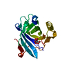
| ||||||||
|---|---|---|---|---|---|---|---|---|---|
| 1 |
| ||||||||
| Unit cell |
|
-Components
| #1: Protein | Mass: 20305.979 Da / Num. of mol.: 1 / Mutation: C40G Source method: isolated from a genetically manipulated source Source: (gene. exp.) Homo sapiens (human) / Gene: 9q34.3 / Cell line (production host): High Five / Production host:  Trichoplusia ni (cabbage looper) / References: UniProt: P07360 Trichoplusia ni (cabbage looper) / References: UniProt: P07360 |
|---|---|
| #2: Chemical | ChemComp-CIT / Citric acid  Mass: 192.124 Da / Num. of mol.: 1 / Source method: obtained synthetically / Formula: C6H8O7 Mass: 192.124 Da / Num. of mol.: 1 / Source method: obtained synthetically / Formula: C6H8O7 |
| #3: Water | ChemComp-HOH / Water Mass: 18.015 Da / Num. of mol.: 186 / Source method: isolated from a natural source / Formula: H2O Mass: 18.015 Da / Num. of mol.: 186 / Source method: isolated from a natural source / Formula: H2O |
-Experimental details
-Experiment
| Experiment | Method: X-RAY DIFFRACTION / Number of used crystals: 1 |
|---|
- Sample preparation
| Crystal | Density Matthews: 2.22 Å3/Da / Density % sol: 44.61 % | ||||||||||||||||||||||||||||||||||||||||||
|---|---|---|---|---|---|---|---|---|---|---|---|---|---|---|---|---|---|---|---|---|---|---|---|---|---|---|---|---|---|---|---|---|---|---|---|---|---|---|---|---|---|---|---|
| Crystal grow | Temperature: 277 K / Method: vapor diffusion, hanging drop / pH: 4 Details: PEG 4000, sodium citrate, pH 4.0, VAPOR DIFFUSION, HANGING DROP, temperature 277K | ||||||||||||||||||||||||||||||||||||||||||
| Crystal grow | *PLUS Temperature: 4 ℃ / pH: 7.2 | ||||||||||||||||||||||||||||||||||||||||||
| Components of the solutions | *PLUS
|
-Data collection
| Diffraction | Mean temperature: 94 K |
|---|---|
| Diffraction source | Source: SYNCHROTRON / Site: APS  / Beamline: 19-ID / Wavelength: 1 Å / Beamline: 19-ID / Wavelength: 1 Å |
| Detector | Type: DECTRIS PILATUS3 X 6M / Detector: PIXEL / Date: Jun 26, 1999 |
| Radiation | Monochromator: High resolution pass with 0.6 degree osc. Low resolution pass with 1.5 degree osc. Protocol: SINGLE WAVELENGTH / Monochromatic (M) / Laue (L): M / Scattering type: x-ray |
| Radiation wavelength | Wavelength: 1 Å / Relative weight: 1 |
| Reflection | Resolution: 1.2→50 Å / Num. all: 57166 / Num. obs: 53030 / % possible obs: 92.2 % / Observed criterion σ(F): 3 / Observed criterion σ(I): 1.5 / Biso Wilson estimate: 9.5 Å2 / Rmerge(I) obs: 0.043 |
| Reflection shell | Resolution: 1.2→1.24 Å / % possible all: 63.3 |
| Reflection | *PLUS Highest resolution: 1.2 Å / Lowest resolution: 50 Å |
| Reflection shell | *PLUS % possible obs: 63.3 % |
- Processing
| Software |
| ||||||||||||||||||||||||||||||||||||
|---|---|---|---|---|---|---|---|---|---|---|---|---|---|---|---|---|---|---|---|---|---|---|---|---|---|---|---|---|---|---|---|---|---|---|---|---|---|
| Refinement | Method to determine structure: MIRAS / Resolution: 1.2→36.03 Å / Rfactor Rfree error: 0.003 / Data cutoff high absF: 364398.04 / Data cutoff low absF: 0 / Isotropic thermal model: RESTRAINED / Cross valid method: THROUGHOUT / σ(F): 0 / Stereochemistry target values: Engh & Huber / Details: The structure was refined also with Turbo Frodo.
| ||||||||||||||||||||||||||||||||||||
| Solvent computation | Solvent model: FLAT MODEL / Bsol: 41.1352 Å2 / ksol: 0.371956 e/Å3 | ||||||||||||||||||||||||||||||||||||
| Displacement parameters | Biso mean: 14.8 Å2
| ||||||||||||||||||||||||||||||||||||
| Refine analyze |
| ||||||||||||||||||||||||||||||||||||
| Refinement step | Cycle: LAST / Resolution: 1.2→36.03 Å
| ||||||||||||||||||||||||||||||||||||
| Refine LS restraints |
| ||||||||||||||||||||||||||||||||||||
| LS refinement shell | Resolution: 1.2→1.28 Å / Rfactor Rfree error: 0.015 / Total num. of bins used: 6
| ||||||||||||||||||||||||||||||||||||
| Xplor file |
|
