 Movie
Movie Controller


+ Open data

- Basic information
| Entry | Database: PDB / ID: 1l9x | ||||||
|---|---|---|---|---|---|---|---|
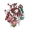
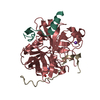
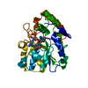
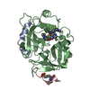
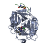
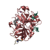
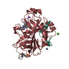
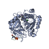
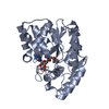
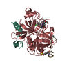
| Title | Structure of gamma-Glutamyl Hydrolase | ||||||
 Components Components | gamma-glutamyl hydrolase | ||||||
 Keywords Keywords | HYDROLASE / gamma-glutamyl hydrolase | ||||||
| Function / homology |  Function and homology information Function and homology informationfolate gamma-glutamyl hydrolase / tetrahydrofolylpolyglutamate metabolic process / gamma-glutamyl-peptidase activity / neutrophil degranulation / exopeptidase activity / response to xenobiotic stimulus => GO:0009410 / vacuole / response to zinc ion / response to insulin / specific granule lumen ...folate gamma-glutamyl hydrolase / tetrahydrofolylpolyglutamate metabolic process / gamma-glutamyl-peptidase activity / neutrophil degranulation / exopeptidase activity / response to xenobiotic stimulus => GO:0009410 / vacuole / response to zinc ion / response to insulin / specific granule lumen / azurophil granule lumen / melanosome / tertiary granule lumen / omega peptidase activity / response to ethanol / Neutrophil degranulation / extracellular space / extracellular exosome / extracellular region / nucleus / cytosolSimilarity search - Function | ||||||
| Biological species |  Homo sapiens (human) Homo sapiens (human) | ||||||
| Method | X-RAY DIFFRACTION / SYNCHROTRON / MAD / Resolution: 1.6 Å | ||||||
 Authors Authors | Li, H. / Ryan, T.J. / Chave, K.J. / Van Roey, P. | ||||||
 Citation Citation | Journal: J.Biol.Chem. / Year: 2002 Title: Three-dimensional structure of human gamma -glutamyl hydrolase. A class I glatamine amidotransferase adapted for a complex substate. Authors: Li, H. / Ryan, T.J. / Chave, K.J. / Van Roey, P. | ||||||
| History |
|
- Structure visualization
| Structure viewer | Molecule: MolmilJmol/JSmol |
|---|
- Downloads & links
-Download
| PDBx/mmCIF format | 1l9x.cif.gz | 256.5 KB | Display | PDBx/mmCIF format |
|---|---|---|---|---|
| PDB format | pdb1l9x.ent.gz | 213.5 KB | Display | PDB format |
| PDBx/mmJSON format | 1l9x.json.gz | Tree view | PDBx/mmJSON format | |
| Others |  Other downloads Other downloads |
-Validation report
| Arichive directory | https://data.pdbj.org/pub/pdb/validation_reports/l9/1l9xftp://data.pdbj.org/pub/pdb/validation_reports/l9/1l9x | HTTPS FTP |
|---|
-Related structure data
| Similar structure data |
|---|
-Links
 PDBj
PDBj


- Assembly
| Deposited unit | 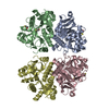
| ||||||||
|---|---|---|---|---|---|---|---|---|---|
| 1 | 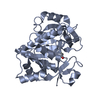
| ||||||||
| 2 | 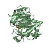
| ||||||||
| 3 | 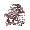
| ||||||||
| 4 | 
| ||||||||
| Unit cell |
|
-Components
| #1: Protein | Mass: 35988.961 Da / Num. of mol.: 4 Source method: isolated from a genetically manipulated source Source: (gene. exp.) Homo sapiens (human) / Plasmid: pET28a / Production host:  Escherichia coli (E. coli) Escherichia coli (E. coli)References: UniProt: Q92820, folate gamma-glutamyl hydrolase #2: Chemical | ChemComp-BME / 2-Mercaptoethanol  Mass: 78.133 Da / Num. of mol.: 6 / Source method: obtained synthetically / Formula: C2H6OS Mass: 78.133 Da / Num. of mol.: 6 / Source method: obtained synthetically / Formula: C2H6OS#3: Water | ChemComp-HOH / | Water Mass: 18.015 Da / Num. of mol.: 820 / Source method: isolated from a natural source / Formula: H2O Mass: 18.015 Da / Num. of mol.: 820 / Source method: isolated from a natural source / Formula: H2O |
|---|
-Experimental details
-Experiment
| Experiment | Method: X-RAY DIFFRACTION / Number of used crystals: 1 |
|---|
- Sample preparation
| Crystal | Density Matthews: 2.58 Å3/Da / Density % sol: 52.24 % | ||||||||||||||||||||||||||||||||||||||||||||||||||||||||||||||||||||||
|---|---|---|---|---|---|---|---|---|---|---|---|---|---|---|---|---|---|---|---|---|---|---|---|---|---|---|---|---|---|---|---|---|---|---|---|---|---|---|---|---|---|---|---|---|---|---|---|---|---|---|---|---|---|---|---|---|---|---|---|---|---|---|---|---|---|---|---|---|---|---|---|
| Crystal grow | Temperature: 288 K / Method: vapor diffusion, hanging drop / pH: 5.6 Details: PEG4000, ammonium acetate, sodium chloride, sodium citrate, pH 5.6, VAPOR DIFFUSION, HANGING DROP, temperature 288.K | ||||||||||||||||||||||||||||||||||||||||||||||||||||||||||||||||||||||
| Crystal grow | *PLUS | ||||||||||||||||||||||||||||||||||||||||||||||||||||||||||||||||||||||
| Components of the solutions | *PLUS
|
-Data collection
| Diffraction | Mean temperature: 100 K | |||||||||||||||
|---|---|---|---|---|---|---|---|---|---|---|---|---|---|---|---|---|
| Diffraction source | Source: SYNCHROTRON / Site: NSLS  / Beamline: X25 / Wavelength: 1.00918, 1.00870, 1.0064, 0.9918 / Beamline: X25 / Wavelength: 1.00918, 1.00870, 1.0064, 0.9918 | |||||||||||||||
| Detector | Type: BRANDEIS - B4 / Detector: CCD / Date: Jun 3, 2001 | |||||||||||||||
| Radiation | Monochromator: GRAPHITE / Protocol: MAD / Monochromatic (M) / Laue (L): M / Scattering type: x-ray | |||||||||||||||
| Radiation wavelength |
| |||||||||||||||
| Reflection | Resolution: 1.6→25 Å / Num. all: 161629 / Num. obs: 161520 / % possible obs: 82.2 % / Observed criterion σ(F): 0 / Observed criterion σ(I): 0 / Redundancy: 6.1 % / Biso Wilson estimate: 21.6 Å2 / Rmerge(I) obs: 0.045 / Rsym value: 0.045 / Net I/σ(I): 53 | |||||||||||||||
| Reflection shell | Resolution: 1.6→1.66 Å / Rmerge(I) obs: 0.211 / Mean I/σ(I) obs: 4.4 / Num. unique all: 6118 / Rsym value: 0.211 / % possible all: 31.5 | |||||||||||||||
| Reflection | *PLUS Redundancy: 6.3 % / Rmerge(I) obs: 0.045 | |||||||||||||||
| Reflection shell | *PLUS % possible obs: 31.5 % / Rmerge(I) obs: 0.211 |
- Processing
| Software |
| |||||||||||||||||||||||||
|---|---|---|---|---|---|---|---|---|---|---|---|---|---|---|---|---|---|---|---|---|---|---|---|---|---|---|
| Refinement | Method to determine structure: MAD / Resolution: 1.6→25 Å / Cross valid method: THROUGHOUT / σ(F): 0 / Stereochemistry target values: Engh & Huber
| |||||||||||||||||||||||||
| Displacement parameters | Biso mean: 24.6 Å2 | |||||||||||||||||||||||||
| Refine analyze | Luzzati coordinate error obs: 0.17 Å / Luzzati sigma a obs: 0.2 Å | |||||||||||||||||||||||||
| Refinement step | Cycle: LAST / Resolution: 1.6→25 Å
| |||||||||||||||||||||||||
| Refine LS restraints |
| |||||||||||||||||||||||||
| Refinement | *PLUS Lowest resolution: 100 Å / Rfactor obs: 0.182 / Rfactor Rfree: 0.202 / Rfactor Rwork: 0.182 | |||||||||||||||||||||||||
| Solvent computation | *PLUS | |||||||||||||||||||||||||
| Displacement parameters | *PLUS | |||||||||||||||||||||||||
| Refine LS restraints | *PLUS
|
