 Movie
Movie Controller

[English] 日本語
 Yorodumi
- PDB-1l0p: CRYSTAL STRUCTURE ANALYSIS OF THE COMPLEX BETWEEN PSYCHROPHILIC A... -
+ Open data

- Basic information
| Entry | Database: PDB / ID: 1l0p | ||||||
|---|---|---|---|---|---|---|---|
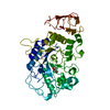
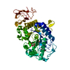
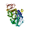
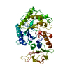
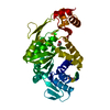
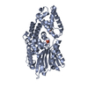
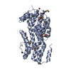
| Title | CRYSTAL STRUCTURE ANALYSIS OF THE COMPLEX BETWEEN PSYCHROPHILIC ALPHA AMYLASE FROM PSEUDOALTEROMONAS HALOPLANCTIS AND NITRATE | ||||||
 Components Components | ALPHA-AMYLASE | ||||||
 Keywords Keywords | HYDROLASE / BETA-ALPHA-8-BARREL / 3 DOMAIN STRUCTURE | ||||||
| Function / homology |  Function and homology informationalpha-amylase / alpha-amylase activity / carbohydrate metabolic process / extracellular region / metal ion binding Function and homology informationalpha-amylase / alpha-amylase activity / carbohydrate metabolic process / extracellular region / metal ion bindingSimilarity search - Function | ||||||
| Biological species |  Pseudoalteromonas haloplanktis (bacteria) Pseudoalteromonas haloplanktis (bacteria) | ||||||
| Method | X-RAY DIFFRACTION / FOURIER SYNTHESIS / Resolution: 2.1 Å | ||||||
 Authors Authors | Aghajari, N. / Haser, R. | ||||||
 Citation Citation | Journal: Protein Sci. / Year: 2002 Title: Structural basis of alpha-amylase activation by chloride. Authors: Aghajari, N. / Feller, G. / Gerday, C. / Haser, R. | ||||||
| History |
|
- Structure visualization
| Structure viewer | Molecule: MolmilJmol/JSmol |
|---|
- Downloads & links
-Download
| PDBx/mmCIF format | 1l0p.cif.gz | 102.8 KB | Display | PDBx/mmCIF format |
|---|---|---|---|---|
| PDB format | pdb1l0p.ent.gz | 77.2 KB | Display | PDB format |
| PDBx/mmJSON format | 1l0p.json.gz | Tree view | PDBx/mmJSON format | |
| Others |  Other downloads Other downloads |
-Validation report
| Arichive directory | https://data.pdbj.org/pub/pdb/validation_reports/l0/1l0pftp://data.pdbj.org/pub/pdb/validation_reports/l0/1l0p | HTTPS FTP |
|---|
-Related structure data
| Related structure data | 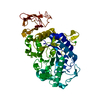 1jd7C  1jd9C 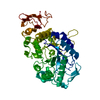 1aqhS S: Starting model for refinement C: citing same article ( |
|---|---|
| Similar structure data |


-Links
 PDBj
PDBj

- Assembly
| Deposited unit | 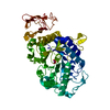
| ||||||||
|---|---|---|---|---|---|---|---|---|---|
| 1 |
| ||||||||
| Unit cell |
|
-Components
| #1: Protein | Mass: 48947.426 Da / Num. of mol.: 1 / Source method: isolated from a natural source / Source: (natural) Pseudoalteromonas haloplanktis (bacteria) / References: UniProt: P29957, alpha-amylase |
|---|---|
| #2: Chemical | ChemComp-CA /   Mass: 40.078 Da / Num. of mol.: 1 / Source method: obtained synthetically / Formula: Ca Mass: 40.078 Da / Num. of mol.: 1 / Source method: obtained synthetically / Formula: Ca |
| #3: Chemical | ChemComp-NO3 / Nitrate  Mass: 62.005 Da / Num. of mol.: 1 / Source method: obtained synthetically / Formula: NO3 Mass: 62.005 Da / Num. of mol.: 1 / Source method: obtained synthetically / Formula: NO3 |
| #4: Water | ChemComp-HOH / Water Mass: 18.015 Da / Num. of mol.: 217 / Source method: isolated from a natural source / Formula: H2O Mass: 18.015 Da / Num. of mol.: 217 / Source method: isolated from a natural source / Formula: H2O |
-Experimental details
-Experiment
| Experiment | Method: X-RAY DIFFRACTION / Number of used crystals: 1 |
|---|
- Sample preparation
| Crystal | Density Matthews: 2.89 Å3/Da / Density % sol: 57.51 % | |||||||||||||||
|---|---|---|---|---|---|---|---|---|---|---|---|---|---|---|---|---|
| Crystal grow | Temperature: 292 K / Method: vapor diffusion, hanging drop / pH: 7 Details: MPD, Hepes, pH 7.00, VAPOR DIFFUSION, HANGING DROP, temperature 292K | |||||||||||||||
| Crystal grow | *PLUS pH: 7 / Details: Aghajari, N., (1996) Protein Sci., 5, 2128. | |||||||||||||||
| Components of the solutions | *PLUS
|
-Data collection
| Diffraction | Mean temperature: 288 K |
|---|---|
| Diffraction source | Source: ROTATING ANODE / Type: ENRAF-NONIUS FR591 / Wavelength: 1.5418 |
| Detector | Type: MARRESEARCH / Detector: IMAGE PLATE / Date: Dec 28, 1998 |
| Radiation | Monochromator: graphite / Protocol: SINGLE WAVELENGTH / Monochromatic (M) / Laue (L): M / Scattering type: x-ray |
| Radiation wavelength | Wavelength: 1.5418 Å / Relative weight: 1 |
| Reflection | Resolution: 2.1→42.6 Å / Num. all: 29937 / Num. obs: 29641 / % possible obs: 89.3 % / Observed criterion σ(I): 1 / Redundancy: 5 % / Biso Wilson estimate: 21.7 Å2 / Rmerge(I) obs: 0.15 |
| Reflection shell | Resolution: 2.1→2.21 Å / Redundancy: 4.5 % / % possible all: 90.5 |
| Reflection | *PLUS Num. measured all: 149687 / Rmerge(I) obs: 0.15 |
| Reflection shell | *PLUS % possible obs: 90.5 % |
- Processing
| Software |
| |||||||||||||||||||||
|---|---|---|---|---|---|---|---|---|---|---|---|---|---|---|---|---|---|---|---|---|---|---|
| Refinement | Method to determine structure: FOURIER SYNTHESIS Starting model: PDB ENTRY 1AQH Resolution: 2.1→42.56 Å / Isotropic thermal model: ISOTROPIC / Cross valid method: THROUGHOUT / σ(F): 0 / Stereochemistry target values: Engh & Huber Details: FINAL CYCLE OF REFINEMENT WAS CARRIED OUT USING CNS.
| |||||||||||||||||||||
| Refinement step | Cycle: LAST / Resolution: 2.1→42.56 Å
| |||||||||||||||||||||
| Refine LS restraints |
| |||||||||||||||||||||
| Refinement | *PLUS Highest resolution: 2.1 Å / Num. reflection obs: 29641 | |||||||||||||||||||||
| Solvent computation | *PLUS | |||||||||||||||||||||
| Displacement parameters | *PLUS |
