 Movie
Movie Controller

[English] 日本語
 Yorodumi
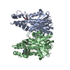
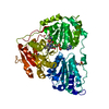
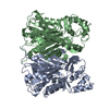
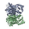
- PDB-1kxt: Camelid VHH Domains in Complex with Porcine Pancreatic alpha-Amylase -
+ Open data

- Basic information
| Entry | Database: PDB / ID: 1kxt | ||||||
|---|---|---|---|---|---|---|---|
| Title | Camelid VHH Domains in Complex with Porcine Pancreatic alpha-Amylase | ||||||
 Components Components |
| ||||||
 Keywords Keywords |  HYDROLASE / IMMUNE SYSTEM / ALPHA 8 BETA 8 / BETA BARREL HYDROLASE / IMMUNE SYSTEM / ALPHA 8 BETA 8 / BETA BARREL | ||||||
| Function / homology |  Function and homology informationalpha-amylase / carbohydrate catabolic process / alpha-amylase activity / chloride ion binding / carbohydrate metabolic process / calcium ion binding / extracellular space Function and homology informationalpha-amylase / carbohydrate catabolic process / alpha-amylase activity / chloride ion binding / carbohydrate metabolic process / calcium ion binding / extracellular spaceSimilarity search - Function | ||||||
| Biological species |  Camelus dromedarius (Arabian camel)Sus scrofa (pig) Camelus dromedarius (Arabian camel)Sus scrofa (pig) | ||||||
| Method | X-RAY DIFFRACTION / SYNCHROTRON / MOLECULAR REPLACEMENT / Resolution: 2 Å | ||||||
 Authors Authors | Desmyter, A. / Spinelli, S. / Payan, F. / Lauwereys, M. / Wyns, L. / Muyldermans, S. / Cambillau, C. | ||||||
 Citation Citation | Journal: J.Biol.Chem. / Year: 2002 Title: Three camelid VHH domains in complex with porcine pancreatic alpha-amylase. Inhibition and versatility of binding topology. Authors: Desmyter, A. / Spinelli, S. / Payan, F. / Lauwereys, M. / Wyns, L. / Muyldermans, S. / Cambillau, C. #1: Journal: Embo J. / Year: 1998Title: Potent enzyme inhibitors derived from dromedary heavy-chain antibodies Authors: Lauwereys, M. / Arbabi Ghahroudi, M. / Desmyter, A. / Kinne, J. / Holzer, W. / De Genst, E. / Wyns, L. / Muyldermans, S. #2: Journal: J.MOL.BIOL. / Year: 1993Title: Structure and Molecular Model Refinement of Pig Pancreatic alpha-amylase at 2.1 A Resolution Authors: Qian, M. / Haser, R. / Payan, F. | ||||||
| History |
| ||||||
| Remark 999 | SEQUENCE The discrepancy between the sequence of this entry and the database reference is explained ...SEQUENCE The discrepancy between the sequence of this entry and the database reference is explained in reference 2 given above. An appropriate sequence database reference for the antibody IMMUNOGLOBULIN VHH FRAGMENT, chains B, D, and F was not available at the time of processing. |
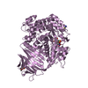
- Structure visualization
| Structure viewer | Molecule: MolmilJmol/JSmol |
|---|
- Downloads & links
-Download
| PDBx/mmCIF format | 1kxt.cif.gz | 400.7 KB | Display | PDBx/mmCIF format |
|---|---|---|---|---|
| PDB format | pdb1kxt.ent.gz | 321.9 KB | Display | PDB format |
| PDBx/mmJSON format | 1kxt.json.gz | Tree view | PDBx/mmJSON format | |
| Others |  Other downloads Other downloads |
-Validation report
| Arichive directory | https://data.pdbj.org/pub/pdb/validation_reports/kx/1kxtftp://data.pdbj.org/pub/pdb/validation_reports/kx/1kxt | HTTPS FTP |
|---|
-Related structure data
| Related structure data | 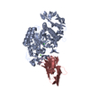 1kxqC 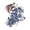 1kxvC  1fjhS  1qdoS C: citing same article ( S: Starting model for refinement |
|---|---|
| Similar structure data |


-Links
 PDBj
PDBj


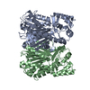
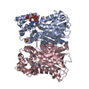
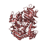
- Assembly
| Deposited unit | 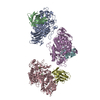
| ||||||||
|---|---|---|---|---|---|---|---|---|---|
| 1 | 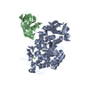
| ||||||||
| 2 | 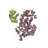
| ||||||||
| 3 | 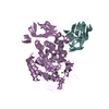
| ||||||||
| Unit cell |
|
-Components
| #1: Protein | / 1 / 4-ALPHA-D-GLUCAN GLUCANOHYDROLASE Mass: 55462.836 Da / Num. of mol.: 3 / Source method: isolated from a natural source / Details: PANCREATIC ENZYME / Source: (natural) Sus scrofa (pig) / References: UniProt: P00690, alpha-amylase#2: Antibody | Mass: 13284.648 Da / Num. of mol.: 3 Source method: isolated from a genetically manipulated source Source: (gene. exp.) Camelus dromedarius (Arabian camel) / Gene: IGG VHH / Plasmid: pHEN6 / Production host:  Escherichia coli (E. coli) / Strain (production host): WK6 Escherichia coli (E. coli) / Strain (production host): WK6#3: Chemical |   Mass: 40.078 Da / Num. of mol.: 3 / Source method: obtained synthetically / Formula: Ca Mass: 40.078 Da / Num. of mol.: 3 / Source method: obtained synthetically / Formula: Ca#4: Chemical | Chloride  Mass: 35.453 Da / Num. of mol.: 3 / Source method: obtained synthetically / Formula: Cl Mass: 35.453 Da / Num. of mol.: 3 / Source method: obtained synthetically / Formula: Cl#5: Water | ChemComp-HOH / | Water Mass: 18.015 Da / Num. of mol.: 1727 / Source method: isolated from a natural source / Formula: H2O Mass: 18.015 Da / Num. of mol.: 1727 / Source method: isolated from a natural source / Formula: H2O |
|---|
-Experimental details
-Experiment
| Experiment | Method: X-RAY DIFFRACTION / Number of used crystals: 1 |
|---|
- Sample preparation
| Crystal | Density Matthews: 2.42 Å3/Da / Density % sol: 49.08 % | |||||||||||||||||||||||||||||||||||
|---|---|---|---|---|---|---|---|---|---|---|---|---|---|---|---|---|---|---|---|---|---|---|---|---|---|---|---|---|---|---|---|---|---|---|---|---|
| Crystal grow | Temperature: 293 K / Method: vapor diffusion, hanging drop / pH: 8 Details: PEG 20000 15%, Malate imidazole 0.2M, pH 8.0, VAPOR DIFFUSION, HANGING DROP, temperature 293K | |||||||||||||||||||||||||||||||||||
| Crystal grow | *PLUS pH: 7.5 | |||||||||||||||||||||||||||||||||||
| Components of the solutions | *PLUS
|
-Data collection
| Diffraction | Mean temperature: 100 K |
|---|---|
| Diffraction source | Source: SYNCHROTRON / Site: ESRF  / Beamline: ID14-2 / Wavelength: 0.98 Å / Beamline: ID14-2 / Wavelength: 0.98 Å |
| Detector | Type: ADSC QUANTUM 4 / Detector: CCD / Date: Nov 20, 2000 / Details: diamond |
| Radiation | Monochromator: diamond / Protocol: SINGLE WAVELENGTH / Monochromatic (M) / Laue (L): M / Scattering type: x-ray |
| Radiation wavelength | Wavelength: 0.98 Å / Relative weight: 1 |
| Reflection | Resolution: 2→39.5 Å / Num. all: 78282 / Num. obs: 65792 / % possible obs: 50 % / Observed criterion σ(F): 0 / Observed criterion σ(I): -3 / Biso Wilson estimate: 7.9 Å2 / Rmerge(I) obs: 0.07 / Net I/σ(I): 7 |
| Reflection shell | Resolution: 2→2.1 Å / Rmerge(I) obs: 0.14 / % possible all: 50 |
| Reflection | *PLUS Lowest resolution: 20 Å / Num. obs: 78282 / % possible obs: 51 % / Num. measured all: 456505 / Rmerge(I) obs: 0.07 |
| Reflection shell | *PLUS Highest resolution: 2 Å / Lowest resolution: 2.13 Å / % possible obs: 50 % / Rmerge(I) obs: 0.137 / Mean I/σ(I) obs: 5.2 |
- Processing
| Software |
| |||||||||||||||||||||||||
|---|---|---|---|---|---|---|---|---|---|---|---|---|---|---|---|---|---|---|---|---|---|---|---|---|---|---|
| Refinement | Method to determine structure: MOLECULAR REPLACEMENT Starting model: PDB ENTRY 1FJH, PDB ENTRY 1QDO Resolution: 2→19.95 Å / Rfactor Rfree error: 0.006 / Data cutoff high absF: 2014619.79 / Data cutoff low absF: 0 / Isotropic thermal model: RESTRAINED / Cross valid method: THROUGHOUT / σ(F): 0 / Stereochemistry target values: Engh & Huber
| |||||||||||||||||||||||||
| Solvent computation | Solvent model: FLAT MODEL / Bsol: 52.8534 Å2 / ksol: 0.37388 e/Å3 | |||||||||||||||||||||||||
| Displacement parameters | Biso mean: 16.9 Å2
| |||||||||||||||||||||||||
| Refine analyze |
| |||||||||||||||||||||||||
| Refinement step | Cycle: LAST / Resolution: 2→19.95 Å
| |||||||||||||||||||||||||
| Refine LS restraints |
| |||||||||||||||||||||||||
| LS refinement shell | Resolution: 2→2.13 Å / Rfactor Rfree error: 0.03 / Total num. of bins used: 6
| |||||||||||||||||||||||||
| Xplor file |
| |||||||||||||||||||||||||
| Refinement | *PLUS Highest resolution: 2 Å / Lowest resolution: 20 Å / Rfactor obs: 0.203 | |||||||||||||||||||||||||
| Solvent computation | *PLUS | |||||||||||||||||||||||||
| Displacement parameters | *PLUS | |||||||||||||||||||||||||
| LS refinement shell | *PLUS Highest resolution: 2 Å / Rfactor obs: 0.228 |
