 Movie
Movie Controller


+ Open data

- Basic information
| Entry | Database: PDB / ID: 1kxo | ||||||
|---|---|---|---|---|---|---|---|
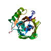
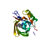
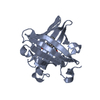
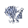
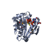
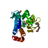
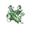
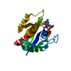
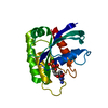
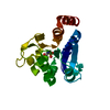
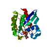
| Title | ENGINEERED LIPOCALIN DIGA16 : APO-FORM | ||||||
 Components Components | DigA16 | ||||||
 Keywords Keywords |  LIGAND BINDING PROTEIN / pieris brassicae / lipocalin / anticalin / genetical engineering / digoxigenin LIGAND BINDING PROTEIN / pieris brassicae / lipocalin / anticalin / genetical engineering / digoxigenin | ||||||
| Function / homology |  Function and homology information Function and homology information | ||||||
| Biological species |  Pieris brassicae (large cabbage white) Pieris brassicae (large cabbage white) | ||||||
| Method | X-RAY DIFFRACTION / FOURIER SYNTHESIS / Resolution: 1.8 Å | ||||||
 Authors Authors | Korndoerfer, I.P. / Skerra, A. | ||||||
 Citation Citation | Journal: J.Mol.Biol. / Year: 2003 Title: Structural mechanism of specific ligand recognition by a lipocalin tailored for the complexation of digoxigenin. Authors: Korndoerfer, I.P. / Schlehuber, S. / Skerra, A. #1: Journal: J.Mol.Biol. / Year: 2000Title: A Novel Type of Receptor Protein, based on the Lipocalin Scaffold, with Specificity for Digoxigenin Authors: Schlehuber, S. / Beste, G. / Skerra, A. #2: Journal: Proc.Natl.Acad.Sci.USA / Year: 1999Title: Small Antibody-like Proteins with Prescribed Ligand Specificities Derived from the Lipocalin Fold. Authors: Beste, G. / Schmidt, F.S. / Stibora, T. / Skerra, A. | ||||||
| History |
|
- Structure visualization
| Structure viewer | Molecule: MolmilJmol/JSmol |
|---|
- Downloads & links
-Download
| PDBx/mmCIF format | 1kxo.cif.gz | 49.3 KB | Display | PDBx/mmCIF format |
|---|---|---|---|---|
| PDB format | pdb1kxo.ent.gz | 35.1 KB | Display | PDB format |
| PDBx/mmJSON format | 1kxo.json.gz | Tree view | PDBx/mmJSON format | |
| Others |  Other downloads Other downloads |
-Validation report
| Arichive directory | https://data.pdbj.org/pub/pdb/validation_reports/kx/1kxoftp://data.pdbj.org/pub/pdb/validation_reports/kx/1kxo | HTTPS FTP |
|---|
-Related structure data


-Links
 PDBj
PDBj

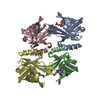
- Assembly
| Deposited unit | 
| ||||||||
|---|---|---|---|---|---|---|---|---|---|
| 1 |
| ||||||||
| Unit cell |
|
-Components
| #1: Protein | Mass: 20833.158 Da / Num. of mol.: 1 Mutation: N1D, N21G, E28Q, K31A, N34D, S35H, V36I, E37T, N48R, H60S, I69S, K87S, L88Y, Y90I, K95Q, N97G, Y114F, K116S, Q125M, F127L, K135M Source method: isolated from a genetically manipulated source Details: genetically engineered variant of bilin binding protein Source: (gene. exp.) Pieris brassicae (large cabbage white) / Plasmid: pbbp21-diga16 / Production host:  Escherichia coli (E. coli) / Strain (production host): jm83 / References: UniProt: P09464 Escherichia coli (E. coli) / Strain (production host): jm83 / References: UniProt: P09464 |
|---|---|
| #2: Water | ChemComp-HOH / Water Mass: 18.015 Da / Num. of mol.: 104 / Source method: isolated from a natural source / Formula: H2O Mass: 18.015 Da / Num. of mol.: 104 / Source method: isolated from a natural source / Formula: H2O |
-Experimental details
-Experiment
| Experiment | Method: X-RAY DIFFRACTION / Number of used crystals: 1 |
|---|
- Sample preparation
| Crystal | Density Matthews: 2.5 Å3/Da / Density % sol: 51.1 % | ||||||||||||||||||||||||||||||||||||
|---|---|---|---|---|---|---|---|---|---|---|---|---|---|---|---|---|---|---|---|---|---|---|---|---|---|---|---|---|---|---|---|---|---|---|---|---|---|
| Crystal grow | Temperature: 298 K / Method: vapor diffusion, hanging drop / pH: 7.6 Details: 0.1-M Hepes, 10% Isopropanole, 19% PEG 4000, pH = 7.8, pH 7.6, VAPOR DIFFUSION, HANGING DROP, temperature 298K | ||||||||||||||||||||||||||||||||||||
| Crystal grow | *PLUS Method: vapor diffusion, hanging drop / PH range low: 8 / PH range high: 7.6 | ||||||||||||||||||||||||||||||||||||
| Components of the solutions | *PLUS
|
-Data collection
| Diffraction | Mean temperature: 298 K |
|---|---|
| Diffraction source | Source: ROTATING ANODE / Type: RIGAKU RU300 / Wavelength: 1.5418 Å |
| Detector | Type: MAR scanner 345 mm plate / Detector: IMAGE PLATE / Date: Nov 6, 2001 |
| Radiation | Monochromator: osmic mirrors / Protocol: SINGLE WAVELENGTH / Monochromatic (M) / Laue (L): M / Scattering type: x-ray |
| Radiation wavelength | Wavelength: 1.5418 Å / Relative weight: 1 |
| Reflection | Resolution: 1.8→48.22 Å / Num. all: 14754 / Num. obs: 14754 / % possible obs: 89.1 % / Observed criterion σ(I): -3 / Redundancy: 2.3 % / Rsym value: 0.04 / Net I/σ(I): 14.2 |
| Reflection shell | Resolution: 1.8→1.86 Å / Redundancy: 1.2 % / Mean I/σ(I) obs: 2.3 / Num. unique all: 1260 / Rsym value: 0.2 / % possible all: 77.2 |
| Reflection | *PLUS Highest resolution: 1.8 Å / Lowest resolution: 99 Å / Num. measured all: 72064 / Rmerge(I) obs: 0.04 |
| Reflection shell | *PLUS % possible obs: 77.2 % / Rmerge(I) obs: 0.2 |
- Processing
| Software |
| ||||||||||||||||||||||||||||||||||||||||||||||||||||||||||||||||||||||||||||||||||||||||||||||||||||||||||||||||||||||||||||||||||
|---|---|---|---|---|---|---|---|---|---|---|---|---|---|---|---|---|---|---|---|---|---|---|---|---|---|---|---|---|---|---|---|---|---|---|---|---|---|---|---|---|---|---|---|---|---|---|---|---|---|---|---|---|---|---|---|---|---|---|---|---|---|---|---|---|---|---|---|---|---|---|---|---|---|---|---|---|---|---|---|---|---|---|---|---|---|---|---|---|---|---|---|---|---|---|---|---|---|---|---|---|---|---|---|---|---|---|---|---|---|---|---|---|---|---|---|---|---|---|---|---|---|---|---|---|---|---|---|---|---|---|---|
| Refinement | Method to determine structure: FOURIER SYNTHESIS Starting model: The structure of diga16 in presence of digoxigenin Resolution: 1.8→48.22 Å / Cor.coef. Fo:Fc: 0.945 / Cor.coef. Fo:Fc free: 0.894 / SU B: 5.518 / SU ML: 0.171 / Cross valid method: THROUGHOUT / σ(F): 0 / ESU R: 0.164 / ESU R Free: 0.176 / Stereochemistry target values: Engh & Huber
| ||||||||||||||||||||||||||||||||||||||||||||||||||||||||||||||||||||||||||||||||||||||||||||||||||||||||||||||||||||||||||||||||||
| Solvent computation | Ion probe radii: 0.8 Å / Shrinkage radii: 0.8 Å / VDW probe radii: 1.4 Å / Solvent model: BABINET MODEL WITH MASK | ||||||||||||||||||||||||||||||||||||||||||||||||||||||||||||||||||||||||||||||||||||||||||||||||||||||||||||||||||||||||||||||||||
| Displacement parameters | Biso mean: 28.316 Å2
| ||||||||||||||||||||||||||||||||||||||||||||||||||||||||||||||||||||||||||||||||||||||||||||||||||||||||||||||||||||||||||||||||||
| Refinement step | Cycle: LAST / Resolution: 1.8→48.22 Å
| ||||||||||||||||||||||||||||||||||||||||||||||||||||||||||||||||||||||||||||||||||||||||||||||||||||||||||||||||||||||||||||||||||
| Refine LS restraints |
| ||||||||||||||||||||||||||||||||||||||||||||||||||||||||||||||||||||||||||||||||||||||||||||||||||||||||||||||||||||||||||||||||||
| LS refinement shell | Resolution: 1.8→1.9 Å / Total num. of bins used: 20
| ||||||||||||||||||||||||||||||||||||||||||||||||||||||||||||||||||||||||||||||||||||||||||||||||||||||||||||||||||||||||||||||||||
| Software | *PLUS Version: 5 / Classification: refinement | ||||||||||||||||||||||||||||||||||||||||||||||||||||||||||||||||||||||||||||||||||||||||||||||||||||||||||||||||||||||||||||||||||
| Refinement | *PLUS Highest resolution: 1.8 Å | ||||||||||||||||||||||||||||||||||||||||||||||||||||||||||||||||||||||||||||||||||||||||||||||||||||||||||||||||||||||||||||||||||
| Solvent computation | *PLUS | ||||||||||||||||||||||||||||||||||||||||||||||||||||||||||||||||||||||||||||||||||||||||||||||||||||||||||||||||||||||||||||||||||
| Displacement parameters | *PLUS | ||||||||||||||||||||||||||||||||||||||||||||||||||||||||||||||||||||||||||||||||||||||||||||||||||||||||||||||||||||||||||||||||||
| Refine LS restraints | *PLUS
|
