 Movie
Movie Controller

[English] 日本語
 Yorodumi
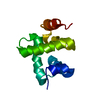
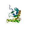
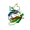
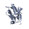
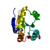
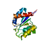
- PDB-1kx9: ANTENNAL CHEMOSENSORY PROTEIN A6 FROM THE MOTH MAMESTRA BRASSICAE -
+ Open data

- Basic information
| Entry | Database: PDB / ID: 1kx9 | ||||||
|---|---|---|---|---|---|---|---|
| Title | ANTENNAL CHEMOSENSORY PROTEIN A6 FROM THE MOTH MAMESTRA BRASSICAE | ||||||
 Components Components | CHEMOSENSORY PROTEIN A6 | ||||||
 Keywords Keywords | LIPID TRANSPORT / ALL HELIX | ||||||
| Function / homology | Antennal chemosensory protein a6 / Insect odorant-binding protein A10/Ejaculatory bulb-specific protein 3 / Insect odorant-binding protein A10/Ejaculatory bulb-specific protein 3 / Insect odorant-binding protein A10/Ejaculatory bulb-specific protein 3 superfamily / Insect pheromone-binding family, A10/OS-D / Orthogonal Bundle / Mainly Alpha /  ACETATE ION / Chemosensory protein ACETATE ION / Chemosensory protein Function and homology information Function and homology information | ||||||
| Biological species |  Mamestra brassicae (cabbage moth) Mamestra brassicae (cabbage moth) | ||||||
| Method | X-RAY DIFFRACTION / SYNCHROTRON / SAD / Resolution: 1.65 Å | ||||||
 Authors Authors | Lartigue, A. / Campanacci, V. / Roussel, A. / Larsson, A.M. / Jones, T.A. / Tegoni, M. / Cambillau, C. | ||||||
 Citation Citation | Journal: J.Biol.Chem. / Year: 2002 Title: X-ray structure and ligand binding study of a moth chemosensory protein Authors: Lartigue, A. / Campanacci, V. / Roussel, A. / Larsson, A.M. / Jones, T.A. / Tegoni, M. / Cambillau, C. #1: Journal: Eur.J.Biochem. / Year: 2001Title: Chemosensory Protein from the Moth Mamestra brassicae. Expression and Secondary Structure from 1H and 15N NMR Authors: Campanacci, V. / Mosbah, A. / Bornet, O. / Wechselberger, R. / Jacquin-Joly, E. / Cambillau, C. / Darbon, H. / Tegoni, M. #2: Journal: Acta Crystallogr.,Sect.D / Year: 2001Title: Recombinant Chemosensory Protein (CSP2) from the moth Mamestra brassicae: Crystallization and Preliminary Crystallographic Study Authors: Campanacci, V. / Spinelli, S. / Lartigue, A. / Lewandowski, C. / Brown, K. / Tegoni, M. / Cambillau, C. | ||||||
| History |
|
- Structure visualization
| Structure viewer | Molecule: MolmilJmol/JSmol |
|---|
- Downloads & links
-Download
| PDBx/mmCIF format | 1kx9.cif.gz | 56.3 KB | Display | PDBx/mmCIF format |
|---|---|---|---|---|
| PDB format | pdb1kx9.ent.gz | 44.6 KB | Display | PDB format |
| PDBx/mmJSON format | 1kx9.json.gz | Tree view | PDBx/mmJSON format | |
| Others |  Other downloads Other downloads |
-Validation report
| Arichive directory | https://data.pdbj.org/pub/pdb/validation_reports/kx/1kx9ftp://data.pdbj.org/pub/pdb/validation_reports/kx/1kx9 | HTTPS FTP |
|---|
-Related structure data


-Links
 PDBj
PDBj- Assembly
| Deposited unit | 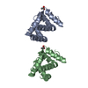
| ||||||||
|---|---|---|---|---|---|---|---|---|---|
| 1 | 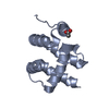
| ||||||||
| 2 | 
| ||||||||
| Unit cell |
|
-Components
| #1: Protein | Mass: 13094.840 Da / Num. of mol.: 2 Source method: isolated from a genetically manipulated source Source: (gene. exp.) Mamestra brassicae (cabbage moth) / Plasmid: pET22b+ / Species (production host): Escherichia coli / Production host:  Escherichia coli BL21 (bacteria) / Strain (production host): BL21 / References: UniProt: Q9NG96 Escherichia coli BL21 (bacteria) / Strain (production host): BL21 / References: UniProt: Q9NG96#2: Chemical | Acetate  Mass: 59.044 Da / Num. of mol.: 2 / Source method: obtained synthetically / Formula: C2H3O2 Mass: 59.044 Da / Num. of mol.: 2 / Source method: obtained synthetically / Formula: C2H3O2#3: Water | ChemComp-HOH / | Water Mass: 18.015 Da / Num. of mol.: 276 / Source method: isolated from a natural source / Formula: H2O Mass: 18.015 Da / Num. of mol.: 276 / Source method: isolated from a natural source / Formula: H2O |
|---|
-Experimental details
-Experiment
| Experiment | Method: X-RAY DIFFRACTION / Number of used crystals: 1 |
|---|
- Sample preparation
| Crystal | Density Matthews: 2.13 Å3/Da / Density % sol: 42.33 % | ||||||||||||||||||||||||||||||
|---|---|---|---|---|---|---|---|---|---|---|---|---|---|---|---|---|---|---|---|---|---|---|---|---|---|---|---|---|---|---|---|
| Crystal grow | Temperature: 293 K / Method: vapor diffusion, hanging drop / pH: 5.5 Details: PEG5000MME 36%, Na Acetate, 0.2M, pH 5.5, VAPOR DIFFUSION, HANGING DROP, temperature 293K | ||||||||||||||||||||||||||||||
| Crystal grow | *PLUS pH: 8 Details: Campanacci, V., (2001) Acta Crystallogr., Sect.D, 57, 137. | ||||||||||||||||||||||||||||||
| Components of the solutions | *PLUS
|
-Data collection
| Diffraction | Mean temperature: 100 K |
|---|---|
| Diffraction source | Source: SYNCHROTRON / Site: ESRF  / Beamline: ID29 / Wavelength: 0.9789 Å / Beamline: ID29 / Wavelength: 0.9789 Å |
| Detector | Type: ADSC QUANTUM 4 / Detector: CCD / Date: Mar 6, 2001 / Details: mirrors |
| Radiation | Protocol: SINGLE WAVELENGTH / Monochromatic (M) / Laue (L): M / Scattering type: x-ray |
| Radiation wavelength | Wavelength: 0.9789 Å / Relative weight: 1 |
| Reflection | Resolution: 1.65→30 Å / Num. all: 25956 / Num. obs: 25956 / % possible obs: 97.6 % / Observed criterion σ(F): 0 / Observed criterion σ(I): 0 / Biso Wilson estimate: 20.5 Å2 / Rmerge(I) obs: 0.038 / Net I/σ(I): 11.8 |
| Reflection shell | Resolution: 1.65→1.75 Å / Rmerge(I) obs: 0.245 / Mean I/σ(I) obs: 7.2 / Num. unique all: 4096 / % possible all: 97.6 |
| Reflection | *PLUS Highest resolution: 1.6 Å / Lowest resolution: 25 Å / % possible obs: 93.3 % |
| Reflection shell | *PLUS Highest resolution: 1.6 Å / Lowest resolution: 1.69 Å |
- Processing
| Software |
| ||||||||||||||||||||||||||||||||||||
|---|---|---|---|---|---|---|---|---|---|---|---|---|---|---|---|---|---|---|---|---|---|---|---|---|---|---|---|---|---|---|---|---|---|---|---|---|---|
| Refinement | Method to determine structure: SAD / Resolution: 1.65→14.93 Å / Rfactor Rfree error: 0.007 / Data cutoff high absF: 1011472.63 / Data cutoff low absF: 0 / Isotropic thermal model: RESTRAINED / Cross valid method: THROUGHOUT / σ(F): 0 / Stereochemistry target values: Engh & Huber
| ||||||||||||||||||||||||||||||||||||
| Solvent computation | Solvent model: FLAT MODEL / Bsol: 51.0333 Å2 / ksol: 0.343558 e/Å3 | ||||||||||||||||||||||||||||||||||||
| Displacement parameters | Biso mean: 19 Å2
| ||||||||||||||||||||||||||||||||||||
| Refine analyze |
| ||||||||||||||||||||||||||||||||||||
| Refinement step | Cycle: LAST / Resolution: 1.65→14.93 Å
| ||||||||||||||||||||||||||||||||||||
| Refine LS restraints |
| ||||||||||||||||||||||||||||||||||||
| LS refinement shell | Resolution: 1.65→1.75 Å / Rfactor Rfree error: 0.019 / Total num. of bins used: 6
| ||||||||||||||||||||||||||||||||||||
| Xplor file |
| ||||||||||||||||||||||||||||||||||||
| Refinement | *PLUS Lowest resolution: 15 Å / Rfactor Rfree: 0.204 / Rfactor Rwork: 0.24 | ||||||||||||||||||||||||||||||||||||
| Solvent computation | *PLUS | ||||||||||||||||||||||||||||||||||||
| Displacement parameters | *PLUS | ||||||||||||||||||||||||||||||||||||
| Refine LS restraints | *PLUS
|
