 Movie
Movie Controller

[English] 日本語
 Yorodumi
- PDB-1ku7: Crystal Structure of Thermus aquatics RNA Polymerase SigmaA Subun... -
+ Open data

- Basic information
| Entry | Database: PDB / ID: 1ku7 | ||||||
|---|---|---|---|---|---|---|---|
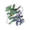
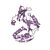
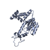
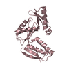
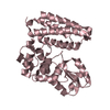
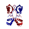
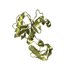
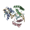
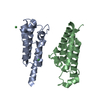
| Title | Crystal Structure of Thermus aquatics RNA Polymerase SigmaA Subunit Region 4 Bound to-35 Element DNA | ||||||
 Components Components |
| ||||||
 Keywords Keywords | TRANSCRIPTION/DNA /  helix-turn-helix / double-helix / TRANSCRIPTION-DNA COMPLEX helix-turn-helix / double-helix / TRANSCRIPTION-DNA COMPLEX | ||||||
| Function / homology |  Function and homology informationsigma factor activity / DNA-templated transcription initiation / DNA binding / cytoplasm Function and homology informationsigma factor activity / DNA-templated transcription initiation / DNA binding / cytoplasmSimilarity search - Function | ||||||
| Biological species |   Thermus aquaticus (bacteria) Thermus aquaticus (bacteria) | ||||||
| Method | X-RAY DIFFRACTION / SYNCHROTRON / MAD / Resolution: 2.4 Å | ||||||
 Authors Authors | Campbell, E.A. / Muzzin, O. / Chlenov, M. / Sun, J.L. / Olson, C.A. / Weinman, O. / Trester-Zedlitz, M.L. / Darst, S.A. | ||||||
 Citation Citation | Journal: Mol.Cell / Year: 2002 Title: Structure of the bacterial RNA polymerase promoter specificity sigma subunit. Authors: Campbell, E.A. / Muzzin, O. / Chlenov, M. / Sun, J.L. / Olson, C.A. / Weinman, O. / Trester-Zedlitz, M.L. / Darst, S.A. | ||||||
| History |
|
- Structure visualization
| Structure viewer | Molecule: MolmilJmol/JSmol |
|---|
- Downloads & links
-Download
| PDBx/mmCIF format | 1ku7.cif.gz | 53.9 KB | Display | PDBx/mmCIF format |
|---|---|---|---|---|
| PDB format | pdb1ku7.ent.gz | 37.6 KB | Display | PDB format |
| PDBx/mmJSON format | 1ku7.json.gz | Tree view | PDBx/mmJSON format | |
| Others |  Other downloads Other downloads |
-Validation report
| Arichive directory | https://data.pdbj.org/pub/pdb/validation_reports/ku/1ku7ftp://data.pdbj.org/pub/pdb/validation_reports/ku/1ku7 | HTTPS FTP |
|---|
-Related structure data


-Links
 PDBj
PDBj


- Assembly
| Deposited unit | 
| ||||||||
|---|---|---|---|---|---|---|---|---|---|
| 1 |
| ||||||||
| Unit cell |
|
-Components
| #1: DNA chain | Mass: 3342.212 Da / Num. of mol.: 1 / Source method: obtained synthetically / Details: -35 Element DNA | ||
|---|---|---|---|
| #2: DNA chain | Mass: 3324.184 Da / Num. of mol.: 1 / Source method: obtained synthetically | ||
| #3: Protein | Mass: 8737.127 Da / Num. of mol.: 2 / Fragment: region 4 (residues 366-438) / Mutation: L386M Source method: isolated from a genetically manipulated source Source: (gene. exp.) Thermus aquaticus (bacteria) / Production host: Escherichia coli (E. coli) / References: UniProt: Q9EZJ8#4: Water | ChemComp-HOH / | Water Mass: 18.015 Da / Num. of mol.: 67 / Source method: isolated from a natural source / Formula: H2O Mass: 18.015 Da / Num. of mol.: 67 / Source method: isolated from a natural source / Formula: H2O |
-Experimental details
-Experiment
| Experiment | Method: X-RAY DIFFRACTION |
|---|
- Sample preparation
| Crystal | Density Matthews: 2.2 Å3/Da / Density % sol: 44.15 % | |||||||||||||||||||||||||||||||||||
|---|---|---|---|---|---|---|---|---|---|---|---|---|---|---|---|---|---|---|---|---|---|---|---|---|---|---|---|---|---|---|---|---|---|---|---|---|
| Crystal grow | Temperature: 298 K / Method: vapor diffusion, hanging drop / pH: 5.5 Details: MES, PEG 4000, magnesium acetate, pH 5.5, VAPOR DIFFUSION, HANGING DROP, temperature 298K | |||||||||||||||||||||||||||||||||||
| Components of the solutions |
| |||||||||||||||||||||||||||||||||||
| Crystal grow | *PLUS Temperature: 22.5 ℃ / pH: 6 / Method: vapor diffusion | |||||||||||||||||||||||||||||||||||
| Components of the solutions | *PLUS
|
-Data collection
| Diffraction | Mean temperature: 93 K |
|---|---|
| Diffraction source | Source: SYNCHROTRON / Site: NSLS  / Beamline: X9A / Wavelength: 0.979 Å / Beamline: X9A / Wavelength: 0.979 Å |
| Detector | Type: MARRESEARCH / Detector: CCD / Date: Aug 9, 2001 |
| Radiation | Monochromator: Si 111 / Protocol: SINGLE WAVELENGTH / Monochromatic (M) / Laue (L): M / Scattering type: x-ray |
| Radiation wavelength | Wavelength: 0.979 Å / Relative weight: 1 |
| Reflection | Resolution: 2.4→25 Å / Num. all: 8898 / Num. obs: 8698 / % possible obs: 97.7 % / Observed criterion σ(F): 0 / Observed criterion σ(I): 0 / Redundancy: 3.8 % |
| Reflection shell | Resolution: 2.4→2.49 Å / Rmerge(I) obs: 0.253 / Mean I/σ(I) obs: 2.9 / % possible all: 79.7 |
| Reflection | *PLUS Highest resolution: 2.4 Å / Lowest resolution: 25 Å / Num. obs: 7801 / Num. measured all: 29108 / Rmerge(I) obs: 0.052 |
| Reflection shell | *PLUS % possible obs: 97.7 % / Rmerge(I) obs: 0.197 / Mean I/σ(I) obs: 5.3 |
- Processing
| Software |
| ||||||||||||||||||||
|---|---|---|---|---|---|---|---|---|---|---|---|---|---|---|---|---|---|---|---|---|---|
| Refinement | Method to determine structure: MAD / Resolution: 2.4→25 Å / Isotropic thermal model: isotropic / Cross valid method: THROUGHOUT / Stereochemistry target values: Engh & Huber
| ||||||||||||||||||||
| Refinement step | Cycle: LAST / Resolution: 2.4→25 Å
| ||||||||||||||||||||
| Refine LS restraints |
| ||||||||||||||||||||
| Refinement | *PLUS Lowest resolution: 25 Å / % reflection Rfree: 5 % | ||||||||||||||||||||
| Solvent computation | *PLUS | ||||||||||||||||||||
| Displacement parameters | *PLUS | ||||||||||||||||||||
| Refine LS restraints | *PLUS
|
