 Movie
Movie Controller

[English] 日本語
 Yorodumi
- PDB-1ks4: The structure of Aspergillus niger endoglucanase-palladium complex -
+ Open data

- Basic information
| Entry | Database: PDB / ID: 1ks4 | ||||||
|---|---|---|---|---|---|---|---|
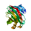
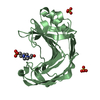
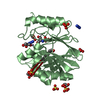
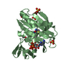
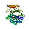
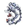
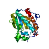
| Title | The structure of Aspergillus niger endoglucanase-palladium complex | ||||||
 Components Components | Endoglucanase A | ||||||
 Keywords Keywords |  HYDROLASE / Endoglucanase / Cellulase / Aspergillus niger / family 9 / (alpha/alpha)6 HYDROLASE / Endoglucanase / Cellulase / Aspergillus niger / family 9 / (alpha/alpha)6 | ||||||
| Function / homology |  Function and homology informationcellulase / cellulase activity / polysaccharide catabolic process / transferase activity Function and homology informationcellulase / cellulase activity / polysaccharide catabolic process / transferase activitySimilarity search - Function | ||||||
| Biological species |  Aspergillus niger (mold) Aspergillus niger (mold) | ||||||
| Method | X-RAY DIFFRACTION / MOLECULAR REPLACEMENT / Resolution: 2.5 Å | ||||||
 Authors Authors | Khademi, S. / Zhang, D. / Swanson, S.M. / Wartenberg, A. / Witte, C. / Meyer, E.F. | ||||||
 Citation Citation | Journal: Acta Crystallogr.,Sect.D / Year: 2002 Title: Determination of the structure of an endoglucanase from Aspergillus niger and its mode of inhibition by palladium chloride. Authors: Khademi, S. / Zhang, D. / Swanson, S.M. / Wartenberg, A. / Witte, K. / Meyer, E.F. | ||||||
| History |
| ||||||
| Remark 650 | HELIX AUTHOR PROVIDED HELIX RECORDS | ||||||
| Remark 700 | SHEET AUTHOR PROVIDED SHEET RECORDS |
- Structure visualization
| Structure viewer | Molecule: MolmilJmol/JSmol |
|---|
- Downloads & links
-Download
| PDBx/mmCIF format | 1ks4.cif.gz | 52.4 KB | Display | PDBx/mmCIF format |
|---|---|---|---|---|
| PDB format | pdb1ks4.ent.gz | 41 KB | Display | PDB format |
| PDBx/mmJSON format | 1ks4.json.gz | Tree view | PDBx/mmJSON format | |
| Others |  Other downloads Other downloads |
-Validation report
| Arichive directory | https://data.pdbj.org/pub/pdb/validation_reports/ks/1ks4ftp://data.pdbj.org/pub/pdb/validation_reports/ks/1ks4 | HTTPS FTP |
|---|
-Related structure data
-Links
 PDBj
PDBj
- Assembly
| Deposited unit | 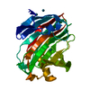
| ||||||||
|---|---|---|---|---|---|---|---|---|---|
| 1 |
| ||||||||
| Unit cell |
|
-Components
| #1: Protein | Mass: 24283.145 Da / Num. of mol.: 1 / Fragment: Aspergillus niger Endoglucanase / Source method: isolated from a natural source / Source: (natural) Aspergillus niger (mold) / Strain: CBS 554.65 / References: UniProt: O74705, cellulase | ||
|---|---|---|---|
| #2: Chemical | Palladium  Mass: 106.420 Da / Num. of mol.: 3 / Source method: obtained synthetically / Formula: Pd Mass: 106.420 Da / Num. of mol.: 3 / Source method: obtained synthetically / Formula: Pd#3: Water | ChemComp-HOH / | Water Mass: 18.015 Da / Num. of mol.: 34 / Source method: isolated from a natural source / Formula: H2O Mass: 18.015 Da / Num. of mol.: 34 / Source method: isolated from a natural source / Formula: H2O |
-Experimental details
-Experiment
| Experiment | Method: X-RAY DIFFRACTION / Number of used crystals: 1 |
|---|
- Sample preparation
| Crystal | Density Matthews: 3.18 Å3/Da / Density % sol: 61.36 % | ||||||||||||||||||||||||
|---|---|---|---|---|---|---|---|---|---|---|---|---|---|---|---|---|---|---|---|---|---|---|---|---|---|
| Crystal grow | Temperature: 291 K / Method: vapor diffusion, hanging drop / pH: 4.8 Details: PEG 4000, Sodium Acetate, pH 4.8, VAPOR DIFFUSION, HANGING DROP, temperature 291K | ||||||||||||||||||||||||
| Crystal grow | *PLUS | ||||||||||||||||||||||||
| Components of the solutions | *PLUS
|
-Data collection
| Diffraction | Mean temperature: 298 K |
|---|---|
| Diffraction source | Source: ROTATING ANODE / Type: RIGAKU RU200 / Wavelength: 1.5418 Å |
| Detector | Type: MAC Science DIP-2030K / Detector: IMAGE PLATE / Date: Oct 1, 1994 / Details: mirrors |
| Radiation | Monochromator: Osmic, Model 140-000023 / Protocol: SINGLE WAVELENGTH / Monochromatic (M) / Laue (L): M / Scattering type: x-ray |
| Radiation wavelength | Wavelength: 1.5418 Å / Relative weight: 1 |
| Reflection | Resolution: 2.5→30 Å / Num. all: 11142 / Num. obs: 10691 / % possible obs: 96 % / Observed criterion σ(F): 1 / Observed criterion σ(I): 3 / Biso Wilson estimate: 22.9 Å2 / Rmerge(I) obs: 0.099 / Net I/σ(I): 26.7 |
| Reflection shell | Resolution: 2.5→2.55 Å / Rmerge(I) obs: 0.09 / Mean I/σ(I) obs: 3.7 / % possible all: 89.5 |
| Reflection | *PLUS Num. obs: 11084 / Num. measured all: 117527 / Rmerge(I) obs: 0.09 |
| Reflection shell | *PLUS Highest resolution: 2.5 Å / % possible obs: 89.3 % / Rmerge(I) obs: 0.63 |
- Processing
| Software |
| ||||||||||||||||||||||||||||||||||||
|---|---|---|---|---|---|---|---|---|---|---|---|---|---|---|---|---|---|---|---|---|---|---|---|---|---|---|---|---|---|---|---|---|---|---|---|---|---|
| Refinement | Method to determine structure: MOLECULAR REPLACEMENT / Resolution: 2.5→30 Å / Rfactor Rfree error: 0.007 / Cross valid method: THROUGHOUT / σ(F): 0 / σ(I): 0 / Stereochemistry target values: Engh & Huber
| ||||||||||||||||||||||||||||||||||||
| Solvent computation | Solvent model: FLAT MODEL / Bsol: 33.743 Å2 / ksol: 0.339 e/Å3 | ||||||||||||||||||||||||||||||||||||
| Displacement parameters | Biso mean: 43.4 Å2
| ||||||||||||||||||||||||||||||||||||
| Refine analyze |
| ||||||||||||||||||||||||||||||||||||
| Refinement step | Cycle: LAST / Resolution: 2.5→30 Å
| ||||||||||||||||||||||||||||||||||||
| Refine LS restraints |
| ||||||||||||||||||||||||||||||||||||
| LS refinement shell | Resolution: 2.5→2.59 Å / Rfactor Rfree error: 0.034 / Total num. of bins used: 10
| ||||||||||||||||||||||||||||||||||||
| Xplor file | Serial no: 1 / Param file: protein_rep.param / Topol file: protein.top | ||||||||||||||||||||||||||||||||||||
| Refinement | *PLUS Rfactor Rfree: 0.242 / Rfactor Rwork: 0.208 | ||||||||||||||||||||||||||||||||||||
| Solvent computation | *PLUS | ||||||||||||||||||||||||||||||||||||
| Displacement parameters | *PLUS | ||||||||||||||||||||||||||||||||||||
| Refine LS restraints | *PLUS
|
