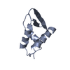
 Movie
Movie Controller


+ Open data

- Basic information
| Entry | Database: PDB / ID: 1kp6 | ||||||
|---|---|---|---|---|---|---|---|
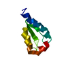
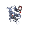
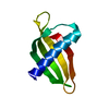
| Title | USTILAGO MAYDIS KILLER TOXIN KP6 ALPHA-SUBUNIT | ||||||
 Components Components | PROTEIN (TOXIN) | ||||||
 Keywords Keywords |  TOXIN / KILLER TOXIN / ION CHANNEL MODULATOR TOXIN / KILLER TOXIN / ION CHANNEL MODULATOR | ||||||
| Function / homology | Killer toxin KP6 alpha-subunit / toxin activity / Alpha-Beta Plaits / 2-Layer Sandwich / extracellular region / Alpha Beta / KP6 killer toxin Function and homology information Function and homology information | ||||||
| Biological species |  Ustilago maydis (fungus) Ustilago maydis (fungus) | ||||||
| Method | X-RAY DIFFRACTION / MIR / Resolution: 1.8 Å | ||||||
 Authors Authors | Li, N. / Erman, M. / Pangborn, W. / Duax, W.L. / Park, C.-M. / Bruenn, J. / Ghosh, D. | ||||||
 Citation Citation | Journal: J.Biol.Chem. / Year: 1999 Title: Structure of Ustilago maydis killer toxin KP6 alpha-subunit. A multimeric assembly with a central pore. Authors: Li, N. / Erman, M. / Pangborn, W. / Duax, W.L. / Park, C.M. / Bruenn, J. / Ghosh, D. | ||||||
| History |
|
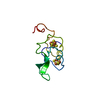
- Structure visualization
| Structure viewer | Molecule: MolmilJmol/JSmol |
|---|
- Downloads & links
-Download
| PDBx/mmCIF format | 1kp6.cif.gz | 33.6 KB | Display | PDBx/mmCIF format |
|---|---|---|---|---|
| PDB format | pdb1kp6.ent.gz | 23 KB | Display | PDB format |
| PDBx/mmJSON format | 1kp6.json.gz | Tree view | PDBx/mmJSON format | |
| Others |  Other downloads Other downloads |
-Validation report
| Arichive directory | https://data.pdbj.org/pub/pdb/validation_reports/kp/1kp6ftp://data.pdbj.org/pub/pdb/validation_reports/kp/1kp6 | HTTPS FTP |
|---|
-Related structure data
| Similar structure data |
|---|


-Links
 PDBj
PDBj- Assembly
| Deposited unit | 
| ||||||||
|---|---|---|---|---|---|---|---|---|---|
| 1 |
| ||||||||
| Unit cell |
|
-Components
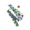
| #1: Protein | Mass: 8736.669 Da / Num. of mol.: 1 / Source method: isolated from a natural source / Source: (natural) Ustilago maydis (fungus) / References: UniProt: P16948 |
|---|---|
| #2: Chemical | ChemComp-SO4 / Sulfate  Mass: 96.063 Da / Num. of mol.: 1 / Source method: obtained synthetically / Formula: SO4 Mass: 96.063 Da / Num. of mol.: 1 / Source method: obtained synthetically / Formula: SO4 |
| #3: Water | ChemComp-HOH / Water Mass: 18.015 Da / Num. of mol.: 77 / Source method: isolated from a natural source / Formula: H2O Mass: 18.015 Da / Num. of mol.: 77 / Source method: isolated from a natural source / Formula: H2O |
-Experimental details
-Experiment
| Experiment | Method: X-RAY DIFFRACTION / Number of used crystals: 1 |
|---|
- Sample preparation
| Crystal | Density Matthews: 2.41 Å3/Da / Density % sol: 42 % | ||||||||||||||||||||||||||||||
|---|---|---|---|---|---|---|---|---|---|---|---|---|---|---|---|---|---|---|---|---|---|---|---|---|---|---|---|---|---|---|---|
| Crystal grow | pH: 6 / Details: 18-21% AM. SULFATE 10MM MES PH 6.0 | ||||||||||||||||||||||||||||||
| Crystal grow | *PLUS Method: vapor diffusion, hanging drop / Details: drop1-0.005 ml, drop2-0.001 ml | ||||||||||||||||||||||||||||||
| Components of the solutions | *PLUS
|
-Data collection
| Diffraction | Mean temperature: 287 K |
|---|---|
| Diffraction source | Source: ROTATING ANODE / Type: RIGAKU RU200 / Wavelength: 1.5418 |
| Detector | Type: RIGAKU RAXIS IIC / Detector: IMAGE PLATE / Date: Sep 1, 1996 |
| Radiation | Monochromator: GRAPHITE / Protocol: SINGLE WAVELENGTH / Monochromatic (M) / Laue (L): M / Scattering type: x-ray |
| Radiation wavelength | Wavelength: 1.5418 Å / Relative weight: 1 |
| Reflection | Resolution: 1.8→99 Å / Num. obs: 8260 / % possible obs: 93.9 % / Redundancy: 13.5 % / Rmerge(I) obs: 0.077 |
| Reflection shell | Resolution: 1.8→1.85 Å / Mean I/σ(I) obs: 2.15 / % possible all: 90 |
| Reflection | *PLUS Num. measured all: 109467 |
| Reflection shell | *PLUS % possible obs: 90 % |
- Processing
| Software |
| ||||||||||||||||||||||||||||||||||||||||||||||||||||||||||||
|---|---|---|---|---|---|---|---|---|---|---|---|---|---|---|---|---|---|---|---|---|---|---|---|---|---|---|---|---|---|---|---|---|---|---|---|---|---|---|---|---|---|---|---|---|---|---|---|---|---|---|---|---|---|---|---|---|---|---|---|---|---|
| Refinement | Method to determine structure: MIR / Resolution: 1.8→8 Å / Cross valid method: THROUGHOUT / σ(F): 2
| ||||||||||||||||||||||||||||||||||||||||||||||||||||||||||||
| Displacement parameters | Biso mean: 17.6 Å2 | ||||||||||||||||||||||||||||||||||||||||||||||||||||||||||||
| Refine analyze | Luzzati coordinate error obs: 0.2 Å | ||||||||||||||||||||||||||||||||||||||||||||||||||||||||||||
| Refinement step | Cycle: LAST / Resolution: 1.8→8 Å
| ||||||||||||||||||||||||||||||||||||||||||||||||||||||||||||
| Refine LS restraints |
| ||||||||||||||||||||||||||||||||||||||||||||||||||||||||||||
| Xplor file | Serial no: 1 / Param file: PARCSDX.PRO / Topol file: TOPHCSDX.PRO |
