 Movie
Movie Controller


+ Open data

- Basic information
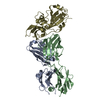
| Entry | Database: PDB / ID: 1kb5 | ||||||
|---|---|---|---|---|---|---|---|
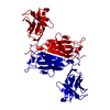
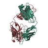
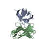
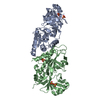
| Title | MURINE T-CELL RECEPTOR VARIABLE DOMAIN/FAB COMPLEX | ||||||
 Components Components |
| ||||||
 Keywords Keywords | COMPLEX (IMMUNOGLOBULIN/RECEPTOR) /  T-CELL RECEPTOR / STRAND SWITCH / FAB / ANTICLONOTYPIC / (IMMUNOGLOBULIN/RECEPTOR) / COMPLEX (IMMUNOGLOBULIN-RECEPTOR) COMPLEX T-CELL RECEPTOR / STRAND SWITCH / FAB / ANTICLONOTYPIC / (IMMUNOGLOBULIN/RECEPTOR) / COMPLEX (IMMUNOGLOBULIN-RECEPTOR) COMPLEX | ||||||
| Function / homology |  Function and homology information Function and homology informationpositive regulation of B cell activation / early endosome to late endosome transport / humoral immune response mediated by circulating immunoglobulin / phagocytosis, recognition / positive regulation of type IIa hypersensitivity / regulation of proteolysis / positive regulation of type I hypersensitivity / antibody-dependent cellular cytotoxicity / Fc-gamma receptor I complex binding / T cell receptor complex ...positive regulation of B cell activation / early endosome to late endosome transport / humoral immune response mediated by circulating immunoglobulin / phagocytosis, recognition / positive regulation of type IIa hypersensitivity / regulation of proteolysis / positive regulation of type I hypersensitivity / antibody-dependent cellular cytotoxicity / Fc-gamma receptor I complex binding / T cell receptor complex / phagocytosis, engulfment / endosome to lysosome transport / positive regulation of endocytosis / immunoglobulin complex, circulating / IgG immunoglobulin complex / immunoglobulin receptor binding / immunoglobulin mediated immune response / antigen processing and presentation / positive regulation of phagocytosis / complement activation, classical pathway / antigen binding / multivesicular body / B cell differentiation / response to bacterium / peptide antigen binding / positive regulation of immune response / antibacterial humoral response / adaptive immune response / extracellular space / extracellular region / plasma membrane / cytosolSimilarity search - Function | ||||||
| Biological species |  Mus musculus (house mouse) Mus musculus (house mouse) | ||||||
| Method | X-RAY DIFFRACTION / SYNCHROTRON / MOLECULAR REPLACEMENT / Resolution: 2.5 Å | ||||||
 Authors Authors | Housset, D. / Mazza, G. / Gregoire, C. / Piras, C. / Malissen, B. / Fontecilla-Camps, J.C. | ||||||
 Citation Citation | Journal: EMBO J. / Year: 1997 Title: The three-dimensional structure of a T-cell antigen receptor V alpha V beta heterodimer reveals a novel arrangement of the V beta domain. Authors: Housset, D. / Mazza, G. / Gregoire, C. / Piras, C. / Malissen, B. / Fontecilla-Camps, J.C. #1: Journal: Science / Year: 1996Title: An Alphabeta T Cell Receptor Structure at 2.5 A and its Orientation in the Tcr-Mhc Complex Authors: Garcia, K.C. / Degano, M. / Stanfield, R.L. / Brunmark, A. / Jackson, M.R. / Peterson, P.A. / Teyton, L. / Wilson, I.A. #2: Journal: Nature / Year: 1996Title: Structure of the Complex between Human T-Cell Receptor, Viral Peptide and Hla-A2 Authors: Garboczi, D.N. / Ghosh, P. / Utz, U. / Fan, Q.R. / Biddison, W.E. / Wiley, D.C. #3: Journal: Science / Year: 1995Title: Crystal Structure of the Beta Chain of a T Cell Antigen Receptor Authors: Bentley, G.A. / Boulot, G. / Karjalainen, K. / Mariuzza, R.A. #4: Journal: Science / Year: 1995Title: Crystal Structure of the V Alpha Domain of a T Cell Antigen Receptor Authors: Fields, B.A. / Ober, B. / Malchiodi, E.L. / Lebedeva, M.I. / Braden, B.C. / Ysern, X. / Kim, J.K. / Shao, X. / Ward, E.S. / Mariuzza, R.A. #5: Journal: Proc.Natl.Acad.Sci.USA / Year: 1991Title: Engineered Secreted T-Cell Receptor Alpha Beta Heterodimers Authors: Gregoire, C. / Rebai, N. / Schweisguth, F. / Necker, A. / Mazza, G. / Auphan, N. / Millward, A. / Schmitt-Verhulst, A.M. / Malissen, B. | ||||||
| History |
|
- Structure visualization
| Structure viewer | Molecule: MolmilJmol/JSmol |
|---|
- Downloads & links
-Download
| PDBx/mmCIF format | 1kb5.cif.gz | 144.1 KB | Display | PDBx/mmCIF format |
|---|---|---|---|---|
| PDB format | pdb1kb5.ent.gz | 114.7 KB | Display | PDB format |
| PDBx/mmJSON format | 1kb5.json.gz | Tree view | PDBx/mmJSON format | |
| Others |  Other downloads Other downloads |
-Validation report
| Arichive directory | https://data.pdbj.org/pub/pdb/validation_reports/kb/1kb5ftp://data.pdbj.org/pub/pdb/validation_reports/kb/1kb5 | HTTPS FTP |
|---|
-Related structure data


-Links
 PDBj
PDBj


- Assembly
| Deposited unit | 
| ||||||||
|---|---|---|---|---|---|---|---|---|---|
| 1 |
| ||||||||
| Unit cell |
|
-Components
| #1: Protein | Mass: 12809.139 Da / Num. of mol.: 1 / Fragment: FV FRAGMENT, VARIABLE DOMAIN Source method: isolated from a genetically manipulated source Details: 24 RESIDUE LINK BETWEEN VALPHA C-TERMINUS AND VBETA N-TERMINUS Source: (gene. exp.) Mus musculus (house mouse) / Cell: T-CELL / Cellular location: SURFACE / Cellular location (production host): MYELOMA / Production host: Mus musculus (house mouse) / References: GenBank: 554285 |
|---|---|
| #2: Protein | Mass: 13381.247 Da / Num. of mol.: 1 / Fragment: FV FRAGMENT, VARIABLE DOMAIN Source method: isolated from a genetically manipulated source Details: 24 RESIDUE LINK BETWEEN VALPHA C-TERMINUS AND VBETA N-TERMINUS Source: (gene. exp.) Mus musculus (house mouse) / Cell: T-CELL / Cellular location: SURFACE / Cellular location (production host): MYELOMA / Production host: Mus musculus (house mouse) / References: UniProt: P04214 |
| #3: Antibody | Mass: 23624.062 Da / Num. of mol.: 1 / Fragment: FAB / Source method: isolated from a natural source / Details: CLEAVED BY PAPAIN / Source: (natural) Mus musculus (house mouse) / Strain: IGG2A / References: UniProt: P01837 |
| #4: Antibody | Mass: 23686.391 Da / Num. of mol.: 1 / Fragment: FAB / Source method: isolated from a natural source / Details: CLEAVED BY PAPAIN / Source: (natural) Mus musculus (house mouse) / Strain: IGG2A / References: UniProt: P01865 |
| #5: Water | ChemComp-HOH / Water Mass: 18.015 Da / Num. of mol.: 266 / Source method: isolated from a natural source / Formula: H2O Mass: 18.015 Da / Num. of mol.: 266 / Source method: isolated from a natural source / Formula: H2O |
| Sequence details | THE TCR FV FRAGMENT AND THE VARIABLE REGIONS OF THE FAB DESIRE-1 HAVE BEEN NUMBERED AS DESCRIBED IN ...THE TCR FV FRAGMENT AND THE VARIABLE REGIONS OF THE FAB DESIRE-1 HAVE BEEN NUMBERED AS DESCRIBED IN KABAT ET AL., 1991. A FEW DISORDERED |
-Experimental details
-Experiment
| Experiment | Method: X-RAY DIFFRACTION / Number of used crystals: 1 |
|---|
- Sample preparation
| Crystal | Density Matthews: 2.5 Å3/Da / Density % sol: 54.18 % | ||||||||||||||||||||||||||||||
|---|---|---|---|---|---|---|---|---|---|---|---|---|---|---|---|---|---|---|---|---|---|---|---|---|---|---|---|---|---|---|---|
| Crystal grow | pH: 7.2 Details: 15% PEG6000, 100MM HEPES PH 6.9-7.5, 200MM NACL, 0.1% NAN3, pH 7.2 PH range: 6.9-7.5 | ||||||||||||||||||||||||||||||
| Crystal | *PLUS | ||||||||||||||||||||||||||||||
| Crystal grow | *PLUS Method: vapor diffusion, hanging drop / PH range low: 7.5 / PH range high: 6.9 | ||||||||||||||||||||||||||||||
| Components of the solutions | *PLUS
|
-Data collection
| Diffraction | Mean temperature: 120 K |
|---|---|
| Diffraction source | Source: SYNCHROTRON / Site: ESRF  / Beamline: BM02 / Wavelength: 0.9786 / Beamline: BM02 / Wavelength: 0.9786 |
| Detector | Detector: CCD / Date: Oct 1, 1996 / Details: MIRRORS |
| Radiation | Monochromator: SI FILTER / Monochromatic (M) / Laue (L): M / Scattering type: x-ray |
| Radiation wavelength | Wavelength: 0.9786 Å / Relative weight: 1 |
| Reflection | Resolution: 2.5→25 Å / Num. obs: 24517 / % possible obs: 86.7 % / Observed criterion σ(I): 0 / Redundancy: 3.1 % / Biso Wilson estimate: 76 Å2 / Rsym value: 0.086 |
| Reflection shell | Resolution: 2.5→2.6 Å / Redundancy: 1.7 % / Rsym value: 0.395 / % possible all: 64.9 |
| Reflection | *PLUS Observed criterion σ(F): 3 / Num. measured all: 75774 / Rmerge(I) obs: 0.086 |
- Processing
| Software |
| ||||||||||||||||||||||||||||||||||||||||||||||||||||||||||||||||||||||||||||||||||||
|---|---|---|---|---|---|---|---|---|---|---|---|---|---|---|---|---|---|---|---|---|---|---|---|---|---|---|---|---|---|---|---|---|---|---|---|---|---|---|---|---|---|---|---|---|---|---|---|---|---|---|---|---|---|---|---|---|---|---|---|---|---|---|---|---|---|---|---|---|---|---|---|---|---|---|---|---|---|---|---|---|---|---|---|---|---|
| Refinement | Method to determine structure: MOLECULAR REPLACEMENT Starting model: PDB ENTRIES 1VFA, 1MLB, 1FLR, 1BEC Resolution: 2.5→10 Å / σ(F): 0 Details: BULK SOLVENT CORRECTION USED R VALUE (WORKING + TEST SET) : 0.221
| ||||||||||||||||||||||||||||||||||||||||||||||||||||||||||||||||||||||||||||||||||||
| Displacement parameters | Biso mean: 48.9 Å2 | ||||||||||||||||||||||||||||||||||||||||||||||||||||||||||||||||||||||||||||||||||||
| Refinement step | Cycle: LAST / Resolution: 2.5→10 Å
| ||||||||||||||||||||||||||||||||||||||||||||||||||||||||||||||||||||||||||||||||||||
| Refine LS restraints |
| ||||||||||||||||||||||||||||||||||||||||||||||||||||||||||||||||||||||||||||||||||||
| Software | *PLUS Name: REFMAC / Classification: refinement | ||||||||||||||||||||||||||||||||||||||||||||||||||||||||||||||||||||||||||||||||||||
| Refinement | *PLUS Num. reflection all: 20232 / Num. reflection obs: 18105 / σ(F): 3 / Rfactor all: 0.221 / Rfactor obs: 0.21 / Rfactor Rfree: 0.315 | ||||||||||||||||||||||||||||||||||||||||||||||||||||||||||||||||||||||||||||||||||||
| Solvent computation | *PLUS | ||||||||||||||||||||||||||||||||||||||||||||||||||||||||||||||||||||||||||||||||||||
| Displacement parameters | *PLUS |
