 Movie
Movie Controller

[English] 日本語
 Yorodumi
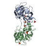
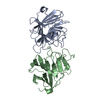
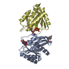
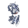
- PDB-1kaq: Structure of Bacillus subtilis Nicotinic Acid Mononucleotide Aden... -
+ Open data

- Basic information
| Entry | Database: PDB / ID: 1kaq | ||||||
|---|---|---|---|---|---|---|---|
| Title | Structure of Bacillus subtilis Nicotinic Acid Mononucleotide Adenylyl Transferase | ||||||
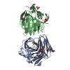
 Components Components | NICOTINATE-NUCLEOTIDE ADENYLYLTRANSFERASE | ||||||
 Keywords Keywords | TRANSFERASE / Rossmann fold / NaAD | ||||||
| Function / homology |  Function and homology informationnicotinate-nucleotide adenylyltransferase / nicotinate-nucleotide adenylyltransferase activity / NAD biosynthetic process / ATP binding Function and homology informationnicotinate-nucleotide adenylyltransferase / nicotinate-nucleotide adenylyltransferase activity / NAD biosynthetic process / ATP bindingSimilarity search - Function | ||||||
| Biological species |  Bacillus subtilis (bacteria) Bacillus subtilis (bacteria) | ||||||
| Method | X-RAY DIFFRACTION / SYNCHROTRON / Combined MAD, molecular replacement phases / Resolution: 3.2 Å | ||||||
 Authors Authors | Olland, A.M. / Underwood, K.W. / Czerwinski, R.M. / Lo, M.C. / Aulabaugh, A. / Bard, J. / Stahl, M.L. / Somers, W.S. / Sullivan, F.X. / Chopra, R. | ||||||
 Citation Citation | Journal: J.Biol.Chem. / Year: 2002 Title: Identification, characterization, and crystal structure of Bacillus subtilis nicotinic acid mononucleotide adenylyltransferase. Authors: Olland, A.M. / Underwood, K.W. / Czerwinski, R.M. / Lo, M.C. / Aulabaugh, A. / Bard, J. / Stahl, M.L. / Somers, W.S. / Sullivan, F.X. / Chopra, R. | ||||||
| History |
|
- Structure visualization
| Structure viewer | Molecule: MolmilJmol/JSmol |
|---|
- Downloads & links
-Download
| PDBx/mmCIF format | 1kaq.cif.gz | 232.2 KB | Display | PDBx/mmCIF format |
|---|---|---|---|---|
| PDB format | pdb1kaq.ent.gz | 190.4 KB | Display | PDB format |
| PDBx/mmJSON format | 1kaq.json.gz | Tree view | PDBx/mmJSON format | |
| Others |  Other downloads Other downloads |
-Validation report
| Arichive directory | https://data.pdbj.org/pub/pdb/validation_reports/ka/1kaqftp://data.pdbj.org/pub/pdb/validation_reports/ka/1kaq | HTTPS FTP |
|---|
-Related structure data


-Links
 PDBj
PDBj- Assembly
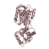
| Deposited unit | 
| ||||||||||
|---|---|---|---|---|---|---|---|---|---|---|---|
| 1 | 
| ||||||||||
| 2 | 
| ||||||||||
| 3 | 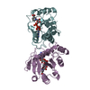
| ||||||||||
| Unit cell |
|
-Components
| #1: Protein | / E.C.2.7.7.18 / DEAMIDO-NAD(+) PYROPHOSPHORYLASE / DEAMIDO-NAD(+) DIPHOSPHORYLASE / NICOTINATE MONONUCLEOTIDE ...DEAMIDO-NAD(+) PYROPHOSPHORYLASE / DEAMIDO-NAD(+) DIPHOSPHORYLASE / NICOTINATE MONONUCLEOTIDE ADENYLYLTRANSFERASE / NAMN ADENYLYLTRANSFERASE / YqeJ Mass: 22618.902 Da / Num. of mol.: 6 Source method: isolated from a genetically manipulated source Source: (gene. exp.) Bacillus subtilis (bacteria) / Gene: nadD / Plasmid: pML208 / Species (production host): Escherichia coli / Production host: Escherichia coli BL21(DE3) (bacteria) / Strain (production host): BL21(DE3)References: UniProt: P54455, nicotinate-nucleotide adenylyltransferase#2: Chemical | ChemComp-DND /   Mass: 665.418 Da / Num. of mol.: 6 / Source method: obtained synthetically / Formula: C21H27N6O15P2 Mass: 665.418 Da / Num. of mol.: 6 / Source method: obtained synthetically / Formula: C21H27N6O15P2 |
|---|
-Experimental details
-Experiment
| Experiment | Method: X-RAY DIFFRACTION / Number of used crystals: 1 |
|---|
- Sample preparation
| Crystal | Density Matthews: 2.78 Å3/Da / Density % sol: 55.83 % | ||||||||||||||||||||||||||||||||||||||||||||||||||||||||
|---|---|---|---|---|---|---|---|---|---|---|---|---|---|---|---|---|---|---|---|---|---|---|---|---|---|---|---|---|---|---|---|---|---|---|---|---|---|---|---|---|---|---|---|---|---|---|---|---|---|---|---|---|---|---|---|---|---|
| Crystal grow | Temperature: 291 K / Method: vapor diffusion, hanging drop / pH: 7.5 Details: PEG 3350, Magnesium Acetate, xylitol, pH 7.5, VAPOR DIFFUSION, HANGING DROP at 291K | ||||||||||||||||||||||||||||||||||||||||||||||||||||||||
| Crystal grow | *PLUS Temperature: 18 ℃ | ||||||||||||||||||||||||||||||||||||||||||||||||||||||||
| Components of the solutions | *PLUS
|
-Data collection
| Diffraction | Mean temperature: 93 K | |||||||||
|---|---|---|---|---|---|---|---|---|---|---|
| Diffraction source | Source: SYNCHROTRON / Site: ALS  / Beamline: 5.0.2 / Wavelength: 0.9791, 0.9639 / Beamline: 5.0.2 / Wavelength: 0.9791, 0.9639 | |||||||||
| Detector | Type: ADSC QUANTUM 4 / Detector: CCD / Date: May 5, 2001 | |||||||||
| Radiation | Monochromator: double crystal / Protocol: MAD / Monochromatic (M) / Laue (L): M / Scattering type: x-ray | |||||||||
| Radiation wavelength |
| |||||||||
| Reflection | Resolution: 3.2→15 Å / Num. all: 25491 / Num. obs: 24599 / % possible obs: 96.5 % / Observed criterion σ(I): 2 | |||||||||
| Reflection shell | Resolution: 3.2→3.31 Å / Rmerge(I) obs: 0.289 / Mean I/σ(I) obs: 5.6 / Num. unique all: 1920 / % possible all: 84 | |||||||||
| Reflection | *PLUS Highest resolution: 3.2 Å / Num. measured all: 80499 / Rmerge(I) obs: 0.147 | |||||||||
| Reflection shell | *PLUS % possible obs: 84 % / Rmerge(I) obs: 0.289 |
- Processing
| Software |
| ||||||||||||||||||||
|---|---|---|---|---|---|---|---|---|---|---|---|---|---|---|---|---|---|---|---|---|---|
| Refinement | Method to determine structure: Combined MAD, molecular replacement phases Resolution: 3.2→15 Å / Cross valid method: THROUGHOUT / σ(F): 2 / Stereochemistry target values: Engh & Huber
| ||||||||||||||||||||
| Refinement step | Cycle: LAST / Resolution: 3.2→15 Å
| ||||||||||||||||||||
| Refine LS restraints |
| ||||||||||||||||||||
| LS refinement shell | Resolution: 3.2→3.23 Å
| ||||||||||||||||||||
| Refinement | *PLUS Highest resolution: 3.2 Å / % reflection Rfree: 5 % / Rfactor Rfree: 0.2975 / Rfactor Rwork: 0.2564 | ||||||||||||||||||||
| Solvent computation | *PLUS | ||||||||||||||||||||
| Displacement parameters | *PLUS | ||||||||||||||||||||
| LS refinement shell | *PLUS Rfactor Rfree: 0.3379 / Rfactor Rwork: 0.3957 |
