 Movie
Movie Controller

[English] 日本語
 Yorodumi
- PDB-1k8t: Crystal structure of the adenylyl cyclase domain of anthrax edema... -
+ Open data

- Basic information
| Entry | Database: PDB / ID: 1k8t | ||||||
|---|---|---|---|---|---|---|---|
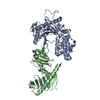
| Title | Crystal structure of the adenylyl cyclase domain of anthrax edema factor (EF) | ||||||
 Components Components | CALMODULIN-SENSITIVE ADENYLATE CYCLASE | ||||||
 Keywords Keywords |  TOXIN / LYASE / edema factor / adenylyl cyclase / anthrax / calmodulin TOXIN / LYASE / edema factor / adenylyl cyclase / anthrax / calmodulin | ||||||
| Function / homology |  Function and homology information Function and homology informationcalcium- and calmodulin-responsive adenylate cyclase activity / calmodulin dependent kinase signaling pathway / adenylate cyclase / cAMP biosynthetic process / adenylate cyclase activity / host cell cytosol / small molecule binding / Uptake and function of anthrax toxins / catalytic complex / metallopeptidase activity ...calcium- and calmodulin-responsive adenylate cyclase activity / calmodulin dependent kinase signaling pathway / adenylate cyclase / cAMP biosynthetic process / adenylate cyclase activity / host cell cytosol / small molecule binding / Uptake and function of anthrax toxins / catalytic complex / metallopeptidase activity / toxin activity / calmodulin binding / extracellular region / ATP binding / metal ion bindingSimilarity search - Function | ||||||
| Biological species |  Bacillus anthracis (anthrax bacterium) Bacillus anthracis (anthrax bacterium) | ||||||
| Method | X-RAY DIFFRACTION / SYNCHROTRON / MOLECULAR REPLACEMENT / Resolution: 2.6 Å | ||||||
 Authors Authors | Drum, C.L. / Yan, S.-Z. / Bard, J. / Shen, Y.-Q. / Lu, D. / Soelaiman, S. / Grabarek, Z. / Bohm, A. / Tang, W.-J. | ||||||
 Citation Citation | Journal: Nature / Year: 2002 Title: Structural basis for the activation of anthrax adenylyl cyclase exotoxin by calmodulin Authors: Drum, C.L. / Yan, S.-Z. / Bard, J. / Shen, Y.-Q. / Lu, D. / Soelaiman, S. / Grabarek, Z. / Bohm, A. / Tang, W.-J. | ||||||
| History |
|
- Structure visualization
| Structure viewer | Molecule: MolmilJmol/JSmol |
|---|
- Downloads & links
-Download
| PDBx/mmCIF format | 1k8t.cif.gz | 115.9 KB | Display | PDBx/mmCIF format |
|---|---|---|---|---|
| PDB format | pdb1k8t.ent.gz | 88.5 KB | Display | PDB format |
| PDBx/mmJSON format | 1k8t.json.gz | Tree view | PDBx/mmJSON format | |
| Others |  Other downloads Other downloads |
-Validation report
| Arichive directory | https://data.pdbj.org/pub/pdb/validation_reports/k8/1k8tftp://data.pdbj.org/pub/pdb/validation_reports/k8/1k8t | HTTPS FTP |
|---|
-Related structure data


-Links
 PDBj
PDBj


- Assembly
| Deposited unit | 
| ||||||||
|---|---|---|---|---|---|---|---|---|---|
| 1 |
| ||||||||
| 2 | 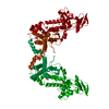
| ||||||||
| Unit cell |
|
-Components
| #1: Protein | Mass: 58810.605 Da / Num. of mol.: 1 Source method: isolated from a genetically manipulated source Source: (gene. exp.) Bacillus anthracis (anthrax bacterium) / Species (production host): Escherichia coli / Production host: Escherichia coli BL21 (bacteria) / Strain (production host): BL21 / References: UniProt: P40136, adenylate cyclase | ||||
|---|---|---|---|---|---|
| #2: Chemical | ChemComp-SO4 / Sulfate  Mass: 96.063 Da / Num. of mol.: 4 / Source method: obtained synthetically / Formula: SO4 Mass: 96.063 Da / Num. of mol.: 4 / Source method: obtained synthetically / Formula: SO4#3: Chemical | ChemComp-NI / | Nickel  Mass: 58.693 Da / Num. of mol.: 1 / Source method: obtained synthetically / Formula: Ni Mass: 58.693 Da / Num. of mol.: 1 / Source method: obtained synthetically / Formula: Ni#4: Water | ChemComp-HOH / | Water Mass: 18.015 Da / Num. of mol.: 96 / Source method: isolated from a natural source / Formula: H2O Mass: 18.015 Da / Num. of mol.: 96 / Source method: isolated from a natural source / Formula: H2O |
-Experimental details
-Experiment
| Experiment | Method: X-RAY DIFFRACTION / Number of used crystals: 1 |
|---|
- Sample preparation
| Crystal | Density Matthews: 3.23 Å3/Da / Density % sol: 61.97 % | |||||||||||||||||||||||||||||||||||||||||||||||||||||||||||||||
|---|---|---|---|---|---|---|---|---|---|---|---|---|---|---|---|---|---|---|---|---|---|---|---|---|---|---|---|---|---|---|---|---|---|---|---|---|---|---|---|---|---|---|---|---|---|---|---|---|---|---|---|---|---|---|---|---|---|---|---|---|---|---|---|---|
| Crystal grow | Temperature: 277 K / Method: vapor diffusion, hanging drop / pH: 5.6 Details: PEG550,Nickle chloride, pH 5.6, VAPOR DIFFUSION, HANGING DROP, temperature 277K | |||||||||||||||||||||||||||||||||||||||||||||||||||||||||||||||
| Crystal grow | *PLUS pH: 7.5 | |||||||||||||||||||||||||||||||||||||||||||||||||||||||||||||||
| Components of the solutions | *PLUS
|
-Data collection
| Diffraction | Mean temperature: 270 K |
|---|---|
| Diffraction source | Source: SYNCHROTRON / Site: APS  / Beamline: 14-BM-D / Wavelength: 1 Å / Beamline: 14-BM-D / Wavelength: 1 Å |
| Detector | Type: ADSC QUANTUM 4 / Detector: CCD |
| Radiation | Protocol: SINGLE WAVELENGTH / Monochromatic (M) / Laue (L): M / Scattering type: x-ray |
| Radiation wavelength | Wavelength: 1 Å / Relative weight: 1 |
| Reflection | Resolution: 2.6→30 Å / Num. obs: 23911 / % possible obs: 98.5 % / Observed criterion σ(F): 1 / Observed criterion σ(I): 2 / Redundancy: 8.7 % / Net I/σ(I): 5.6 |
| Reflection shell | Highest resolution: 2.6 Å / Rmerge(I) obs: 0.273 / Mean I/σ(I) obs: 1.7 / % possible all: 95.4 |
| Reflection | *PLUS Lowest resolution: 30 Å / Redundancy: 6.5 % / Rmerge(I) obs: 0.087 |
| Reflection shell | *PLUS % possible obs: 95.4 % / Redundancy: 4.8 % |
- Processing
| Software |
| |||||||||||||||||||||
|---|---|---|---|---|---|---|---|---|---|---|---|---|---|---|---|---|---|---|---|---|---|---|
| Refinement | Method to determine structure: MOLECULAR REPLACEMENT Starting model: EF-CaM Resolution: 2.6→30 Å / Cross valid method: THROUGHOUT / σ(F): 0 / σ(I): 0 / Stereochemistry target values: Engh & Huber
| |||||||||||||||||||||
| Displacement parameters | Biso mean: 58.8 Å2
| |||||||||||||||||||||
| Refine analyze | Luzzati coordinate error obs: 0.33 Å / Luzzati d res low obs: 5 Å / Luzzati sigma a obs: 0.26 Å | |||||||||||||||||||||
| Refinement step | Cycle: LAST / Resolution: 2.6→30 Å
| |||||||||||||||||||||
| Refine LS restraints |
| |||||||||||||||||||||
| Software | *PLUS Name: CNS / Version: 1 / Classification: refinement | |||||||||||||||||||||
| Refinement | *PLUS Lowest resolution: 30 Å / σ(F): 0 / Rfactor obs: 0.228 | |||||||||||||||||||||
| Solvent computation | *PLUS | |||||||||||||||||||||
| Displacement parameters | *PLUS Biso mean: 58.8 Å2 | |||||||||||||||||||||
| Refine LS restraints | *PLUS
|
