 Movie
Movie Controller

[English] 日本語
 Yorodumi
- PDB-1k6v: LACK OF SYNERGY FOR INHIBITORS TARGETING A MULTI-DRUG RESISTANT H... -
+ Open data

- Basic information
| Entry | Database: PDB / ID: 1k6v | ||||||
|---|---|---|---|---|---|---|---|
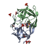
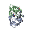
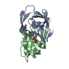
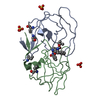
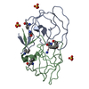
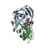
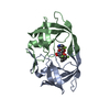
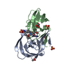
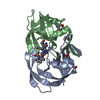
| Title | LACK OF SYNERGY FOR INHIBITORS TARGETING A MULTI-DRUG RESISTANT HIV-1 PROTEASE | ||||||
 Components Components | POL polyprotein | ||||||
 Keywords Keywords |  HYDROLASE / INDINAVIR / INHIBITOR RECOGNITION / DRUG RESISTANCE / HIV-1 PROTEASE HYDROLASE / INDINAVIR / INHIBITOR RECOGNITION / DRUG RESISTANCE / HIV-1 PROTEASE | ||||||
| Function / homology |  Function and homology informationHIV-1 retropepsin / : / retroviral ribonuclease H / exoribonuclease H / : / exoribonuclease H activity / host multivesicular body / DNA integration / RNA-directed DNA polymerase / viral genome integration into host DNA ...HIV-1 retropepsin / : / retroviral ribonuclease H / exoribonuclease H / : / exoribonuclease H activity / host multivesicular body / DNA integration / RNA-directed DNA polymerase / viral genome integration into host DNA / viral penetration into host nucleus / establishment of integrated proviral latency / RNA-directed DNA polymerase activity / Transferases; Transferring phosphorus-containing groups; Nucleotidyltransferases / RNA-DNA hybrid ribonuclease activity / viral nucleocapsid / DNA recombination / Hydrolases; Acting on ester bonds / DNA-directed DNA polymerase / aspartic-type endopeptidase activity / DNA-directed DNA polymerase activity / symbiont entry into host cell / symbiont-mediated suppression of host gene expression / lipid binding / host cell nucleus / structural molecule activity / host cell plasma membrane / virion membrane / proteolysis / DNA binding / RNA binding / zinc ion binding / membrane Function and homology informationHIV-1 retropepsin / : / retroviral ribonuclease H / exoribonuclease H / : / exoribonuclease H activity / host multivesicular body / DNA integration / RNA-directed DNA polymerase / viral genome integration into host DNA ...HIV-1 retropepsin / : / retroviral ribonuclease H / exoribonuclease H / : / exoribonuclease H activity / host multivesicular body / DNA integration / RNA-directed DNA polymerase / viral genome integration into host DNA / viral penetration into host nucleus / establishment of integrated proviral latency / RNA-directed DNA polymerase activity / Transferases; Transferring phosphorus-containing groups; Nucleotidyltransferases / RNA-DNA hybrid ribonuclease activity / viral nucleocapsid / DNA recombination / Hydrolases; Acting on ester bonds / DNA-directed DNA polymerase / aspartic-type endopeptidase activity / DNA-directed DNA polymerase activity / symbiont entry into host cell / symbiont-mediated suppression of host gene expression / lipid binding / host cell nucleus / structural molecule activity / host cell plasma membrane / virion membrane / proteolysis / DNA binding / RNA binding / zinc ion binding / membraneSimilarity search - Function | ||||||
| Biological species |   Human immunodeficiency virus 1 Human immunodeficiency virus 1 | ||||||
| Method | X-RAY DIFFRACTION / FOURIER SYNTHESIS / Resolution: 2 Å | ||||||
 Authors Authors | Schiffer, C.A. | ||||||
 Citation Citation | Journal: Protein Sci. / Year: 2002 Title: Lack of synergy for inhibitors targeting a multi-drug-resistant HIV-1 protease. Authors: King, N.M. / Melnick, L. / Prabu-Jeyabalan, M. / Nalivaika, E.A. / Yang, S.S. / Gao, Y. / Nie, X. / Zepp, C. / Heefner, D.L. / Schiffer, C.A. | ||||||
| History |
|
- Structure visualization
| Structure viewer | Molecule: MolmilJmol/JSmol |
|---|
- Downloads & links
-Download
| PDBx/mmCIF format | 1k6v.cif.gz | 58.2 KB | Display | PDBx/mmCIF format |
|---|---|---|---|---|
| PDB format | pdb1k6v.ent.gz | 41.5 KB | Display | PDB format |
| PDBx/mmJSON format | 1k6v.json.gz | Tree view | PDBx/mmJSON format | |
| Others |  Other downloads Other downloads |
-Validation report
| Arichive directory | https://data.pdbj.org/pub/pdb/validation_reports/k6/1k6vftp://data.pdbj.org/pub/pdb/validation_reports/k6/1k6v | HTTPS FTP |
|---|
-Related structure data
| Related structure data |  1k6cC  1k6pSC  1k6tC C: citing same article ( S: Starting model for refinement |
|---|---|
| Similar structure data |


-Links
 PDBj
PDBj

- Assembly
| Deposited unit | 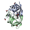
| ||||||||
|---|---|---|---|---|---|---|---|---|---|
| 1 |
| ||||||||
| Unit cell |
|
-Components
| #1: Protein | Mass: 10831.750 Da / Num. of mol.: 2 / Fragment: HIV-1 PROTEASE, Residues 57-155 / Mutation: Q7K,K14R,V82T,I84V Source method: isolated from a genetically manipulated source Source: (gene. exp.) Human immunodeficiency virus 1 / Genus: Lentivirus / Gene: POL / Production host:  Escherichia coli (E. coli) / References: UniProt: P35963, HIV-1 retropepsin Escherichia coli (E. coli) / References: UniProt: P35963, HIV-1 retropepsin#2: Chemical | ChemComp-ACT / Acetate  Mass: 59.044 Da / Num. of mol.: 4 / Source method: obtained synthetically / Formula: C2H3O2 Mass: 59.044 Da / Num. of mol.: 4 / Source method: obtained synthetically / Formula: C2H3O2#3: Chemical | ChemComp-XN2 / |   Mass: 670.837 Da / Num. of mol.: 1 / Source method: obtained synthetically / Formula: C39H50N4O6 Mass: 670.837 Da / Num. of mol.: 1 / Source method: obtained synthetically / Formula: C39H50N4O6#4: Water | ChemComp-HOH / | Water Mass: 18.015 Da / Num. of mol.: 158 / Source method: isolated from a natural source / Formula: H2O Mass: 18.015 Da / Num. of mol.: 158 / Source method: isolated from a natural source / Formula: H2O |
|---|
-Experimental details
-Experiment
| Experiment | Method: X-RAY DIFFRACTION / Number of used crystals: 1 |
|---|
- Sample preparation
| Crystal | Density Matthews: 2.18 Å3/Da / Density % sol: 43.48 % | ||||||||||||||||||||||||||||||||||||||||||||||||||||||
|---|---|---|---|---|---|---|---|---|---|---|---|---|---|---|---|---|---|---|---|---|---|---|---|---|---|---|---|---|---|---|---|---|---|---|---|---|---|---|---|---|---|---|---|---|---|---|---|---|---|---|---|---|---|---|---|
| Crystal grow | Temperature: 298 K / Method: vapor diffusion, hanging drop / pH: 6.2 Details: SODIUM PHOSPHATE, SODIUM CITRATE, AMMONIUM SULPHATE, pH 6.2, VAPOR DIFFUSION, HANGING DROP, temperature 298K | ||||||||||||||||||||||||||||||||||||||||||||||||||||||
| Crystal grow | *PLUS pH: 5.5 | ||||||||||||||||||||||||||||||||||||||||||||||||||||||
| Components of the solutions | *PLUS
|
-Data collection
| Diffraction | Mean temperature: 298 K |
|---|---|
| Diffraction source | Source: ROTATING ANODE / Type: RIGAKU / Wavelength: 1.5418 / Wavelength: 1.5418 Å |
| Detector | Type: RIGAKU RAXIS IV / Detector: IMAGE PLATE / Date: Apr 11, 2000 / Details: MIRRORS |
| Radiation | Monochromator: YALE MIRRORS / Protocol: SINGLE WAVELENGTH / Monochromatic (M) / Laue (L): M / Scattering type: x-ray |
| Radiation wavelength | Wavelength: 1.5418 Å / Relative weight: 1 |
| Reflection | Resolution: 2→27 Å / Num. all: 12820 / Num. obs: 12820 / % possible obs: 96 % / Observed criterion σ(F): 0 / Observed criterion σ(I): 0 / Redundancy: 8.5 % / Rmerge(I) obs: 0.069 / Net I/σ(I): 8.6 |
| Reflection | *PLUS Lowest resolution: 27 Å / Num. measured all: 95068 |
- Processing
| Software |
| ||||||||||||||||||||||||||||||||||||||||||||||||||||||||||||
|---|---|---|---|---|---|---|---|---|---|---|---|---|---|---|---|---|---|---|---|---|---|---|---|---|---|---|---|---|---|---|---|---|---|---|---|---|---|---|---|---|---|---|---|---|---|---|---|---|---|---|---|---|---|---|---|---|---|---|---|---|---|
| Refinement | Method to determine structure: FOURIER SYNTHESIS Starting model: PDB ENTRY 1K6P Resolution: 2→27 Å / Isotropic thermal model: RESTRAINED / Cross valid method: THROUGHOUT / σ(F): 0 / Stereochemistry target values: ENGH & HUBER
| ||||||||||||||||||||||||||||||||||||||||||||||||||||||||||||
| Refinement step | Cycle: LAST / Resolution: 2→27 Å
| ||||||||||||||||||||||||||||||||||||||||||||||||||||||||||||
| Refine LS restraints |
| ||||||||||||||||||||||||||||||||||||||||||||||||||||||||||||
| Xplor file |
| ||||||||||||||||||||||||||||||||||||||||||||||||||||||||||||
| Software | *PLUS Name: CNS / Version: 0.9 / Classification: refinement | ||||||||||||||||||||||||||||||||||||||||||||||||||||||||||||
| Refinement | *PLUS Lowest resolution: 27 Å / % reflection Rfree: 10 % | ||||||||||||||||||||||||||||||||||||||||||||||||||||||||||||
| Solvent computation | *PLUS | ||||||||||||||||||||||||||||||||||||||||||||||||||||||||||||
| Displacement parameters | *PLUS | ||||||||||||||||||||||||||||||||||||||||||||||||||||||||||||
| Refine LS restraints | *PLUS
|
