 Movie
Movie Controller

[English] 日本語
 Yorodumi
- PDB-1k4j: Crystal Structure of the Acyl-homoserinelactone Synthase EsaI Com... -
+ Open data

- Basic information
| Entry | Database: PDB / ID: 1k4j | ||||||
|---|---|---|---|---|---|---|---|
| Title | Crystal Structure of the Acyl-homoserinelactone Synthase EsaI Complexed with Rhenate | ||||||
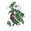
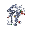
 Components Components | acyl-homoserinelactone synthase EsaI | ||||||
 Keywords Keywords |  LIGASE / acyl-homoserinelactone synthase / mixed alpha beta / enzyme / GNAT similarity / quorum sensing / autoinducer synthase LIGASE / acyl-homoserinelactone synthase / mixed alpha beta / enzyme / GNAT similarity / quorum sensing / autoinducer synthase | ||||||
| Function / homology |  Function and homology informationacyl-homoserine-lactone synthase / N-acyl homoserine lactone synthase activity / quorum sensing Function and homology informationacyl-homoserine-lactone synthase / N-acyl homoserine lactone synthase activity / quorum sensingSimilarity search - Function | ||||||
| Biological species |  Pantoea stewartii subsp. stewartii (bacteria) Pantoea stewartii subsp. stewartii (bacteria) | ||||||
| Method | X-RAY DIFFRACTION / SYNCHROTRON / MAD / Resolution: 2.5 Å | ||||||
 Authors Authors | Watson, W.T. / Minogue, T.D. / Val, D.L. / Beck von Bodman, S. / Churchill, M.E.A. | ||||||
 Citation Citation | Journal: Mol.Cell / Year: 2002 Title: Structural basis and specificity of acyl-homoserine lactone signal production in bacterial quorum sensing. Authors: Watson, W.T. / Minogue, T.D. / Val, D.L. / von Bodman, S.B. / Churchill, M.E. #1: Journal: Acta Crystallogr.,Sect.D / Year: 2001Title: Crystallization and Rhenium MAD Phasing of the Acyl-homoserinelactone Synthase EsaI Authors: Watson, W.T. / Murphy, F.V. / Gould, T.A. / Jambeck, P. / Val, D.L. / Cronan, J.E. / Beck von Bodman, S. / Churchill, M.E.A. | ||||||
| History |
|
- Structure visualization
| Structure viewer | Molecule: MolmilJmol/JSmol |
|---|
- Downloads & links
-Download
| PDBx/mmCIF format | 1k4j.cif.gz | 52.7 KB | Display | PDBx/mmCIF format |
|---|---|---|---|---|
| PDB format | pdb1k4j.ent.gz | 37.5 KB | Display | PDB format |
| PDBx/mmJSON format | 1k4j.json.gz | Tree view | PDBx/mmJSON format | |
| Others |  Other downloads Other downloads |
-Validation report
| Arichive directory | https://data.pdbj.org/pub/pdb/validation_reports/k4/1k4jftp://data.pdbj.org/pub/pdb/validation_reports/k4/1k4j | HTTPS FTP |
|---|
-Related structure data


-Links
 PDBj
PDBj

- Assembly
| Deposited unit | 
| ||||||||
|---|---|---|---|---|---|---|---|---|---|
| 1 |
| ||||||||
| Unit cell |
|
-Components
| #1: Protein | Mass: 26040.727 Da / Num. of mol.: 1 Source method: isolated from a genetically manipulated source Source: (gene. exp.) Pantoea stewartii subsp. stewartii (bacteria)Species: Pantoea stewartii / Strain: subsp. stewartii / Gene: esaI/esaR cluster / Plasmid: pET14b / Species (production host): Escherichia coli / Production host: Escherichia coli BL21(DE3) (bacteria) / Strain (production host): BL21(DE3) / References: UniProt: P54656 | ||
|---|---|---|---|
| #2: Chemical | ChemComp-REO / Perrhenate  Mass: 250.205 Da / Num. of mol.: 7 / Source method: obtained synthetically / Formula: O4Re Mass: 250.205 Da / Num. of mol.: 7 / Source method: obtained synthetically / Formula: O4Re#3: Water | ChemComp-HOH / | Water Mass: 18.015 Da / Num. of mol.: 21 / Source method: isolated from a natural source / Formula: H2O Mass: 18.015 Da / Num. of mol.: 21 / Source method: isolated from a natural source / Formula: H2O |
-Experimental details
-Experiment
| Experiment | Method: X-RAY DIFFRACTION / Number of used crystals: 1 |
|---|
- Sample preparation
| Crystal | Density Matthews: 2 Å3/Da / Density % sol: 38.55 % | ||||||||||||||||||||||||||||||||||||||||||||||||||||||||
|---|---|---|---|---|---|---|---|---|---|---|---|---|---|---|---|---|---|---|---|---|---|---|---|---|---|---|---|---|---|---|---|---|---|---|---|---|---|---|---|---|---|---|---|---|---|---|---|---|---|---|---|---|---|---|---|---|---|
| Crystal grow | Temperature: 291 K / Method: vapor diffusion, hanging drop / pH: 6.1 Details: PEG 4000, 2-propanol, MES, EDTA, beta-mercaptoethanol, NaN3, pH 6.1, VAPOR DIFFUSION, HANGING DROP, temperature 291K | ||||||||||||||||||||||||||||||||||||||||||||||||||||||||
| Crystal grow | *PLUS Method: vapor diffusion | ||||||||||||||||||||||||||||||||||||||||||||||||||||||||
| Components of the solutions | *PLUS
|
-Data collection
| Diffraction | Mean temperature: 113 K | |||||||||||||||
|---|---|---|---|---|---|---|---|---|---|---|---|---|---|---|---|---|
| Diffraction source | Source: SYNCHROTRON / Site: NSLS  / Beamline: X12C / Wavelength: 1.14, 1.1724, 1.1719, 1.19 / Beamline: X12C / Wavelength: 1.14, 1.1724, 1.1719, 1.19 | |||||||||||||||
| Detector | Type: BRANDEIS - B4 / Detector: CCD / Date: Nov 13, 1999 / Details: monochromator | |||||||||||||||
| Radiation | Monochromator: parallel silicon crystal pair / Protocol: MAD / Monochromatic (M) / Laue (L): M / Scattering type: x-ray | |||||||||||||||
| Radiation wavelength |
| |||||||||||||||
| Reflection | Resolution: 2.5→30 Å / Num. all: 7168 / Num. obs: 7101 / % possible obs: 97 % / Observed criterion σ(F): 4 / Observed criterion σ(I): 7.3 / Redundancy: 4.5 % / Biso Wilson estimate: 54.4 Å2 / Rsym value: 0.042 / Net I/σ(I): 20.9 | |||||||||||||||
| Reflection shell | Resolution: 2.5→2.6 Å / Redundancy: 2.4 % / Mean I/σ(I) obs: 3.2 / Num. unique all: 1373 / Rsym value: 0.175 / % possible all: 98.1 | |||||||||||||||
| Reflection | *PLUS Lowest resolution: 30 Å / Num. obs: 7283 / Num. measured all: 32922 / Rmerge(I) obs: 0.042 | |||||||||||||||
| Reflection shell | *PLUS % possible obs: 98.1 % / Rmerge(I) obs: 0.175 / Mean I/σ(I) obs: 4.7 |
- Processing
| Software |
| |||||||||||||||||||||||||
|---|---|---|---|---|---|---|---|---|---|---|---|---|---|---|---|---|---|---|---|---|---|---|---|---|---|---|
| Refinement | Method to determine structure: MAD Starting model: none Resolution: 2.5→30 Å / Isotropic thermal model: RESTRAINED / Cross valid method: THROUGHOUT / σ(F): 2 / σ(I): 0 / Stereochemistry target values: Engh & Huber
| |||||||||||||||||||||||||
| Displacement parameters | Biso mean: 50.9 Å2
| |||||||||||||||||||||||||
| Refine analyze |
| |||||||||||||||||||||||||
| Refinement step | Cycle: LAST / Resolution: 2.5→30 Å
| |||||||||||||||||||||||||
| Refine LS restraints |
| |||||||||||||||||||||||||
| LS refinement shell | Resolution: 2.5→2.59 Å / Rfactor Rfree error: 0.038
| |||||||||||||||||||||||||
| Refinement | *PLUS % reflection Rfree: 11 % / Rfactor all: 0.222 / Rfactor obs: 0.216 / Rfactor Rfree: 0.275 / Rfactor Rwork: 0.216 | |||||||||||||||||||||||||
| Solvent computation | *PLUS | |||||||||||||||||||||||||
| Displacement parameters | *PLUS | |||||||||||||||||||||||||
| Refine LS restraints | *PLUS
| |||||||||||||||||||||||||
| LS refinement shell | *PLUS Highest resolution: 2.5 Å / Rfactor Rfree: 0.291 / Rfactor Rwork: 0.253 |
