 Movie
Movie Controller

[English] 日本語
 Yorodumi
- PDB-1k3b: Crystal Structure of Human Dipeptidyl Peptidase I (Cathepsin C): ... -
+ Open data

- Basic information
| Entry | Database: PDB / ID: 1k3b | ||||||
|---|---|---|---|---|---|---|---|
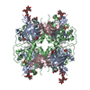
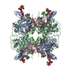
| Title | Crystal Structure of Human Dipeptidyl Peptidase I (Cathepsin C): Exclusion Domain Added to an Endopeptidase Framework Creates the Machine for Activation of Granular Serine Proteases | ||||||
 Components Components | (dipeptydil-peptidase I ...) x 3 | ||||||
 Keywords Keywords |  HYDROLASE HYDROLASE | ||||||
| Function / homology |  Function and homology informationdipeptidyl-peptidase I / peptidase activator activity involved in apoptotic process / positive regulation of proteolysis involved in protein catabolic process / negative regulation of myelination / positive regulation of microglial cell activation / Cargo concentration in the ER / dipeptidyl-peptidase activity / COPII-mediated vesicle transport / chloride ion binding / COPII-coated ER to Golgi transport vesicle ...dipeptidyl-peptidase I / peptidase activator activity involved in apoptotic process / positive regulation of proteolysis involved in protein catabolic process / negative regulation of myelination / positive regulation of microglial cell activation / Cargo concentration in the ER / dipeptidyl-peptidase activity / COPII-mediated vesicle transport / chloride ion binding / COPII-coated ER to Golgi transport vesicle / phosphatase binding / cysteine-type peptidase activity / positive regulation of apoptotic signaling pathway / MHC class II antigen presentation / endoplasmic reticulum-Golgi intermediate compartment membrane / proteolysis involved in protein catabolic process / response to organic substance / T cell mediated cytotoxicity / : / azurophil granule lumen / protein-folding chaperone binding / collagen-containing extracellular matrix / lysosome / immune response / endoplasmic reticulum lumen / cysteine-type endopeptidase activity / serine-type endopeptidase activity / intracellular membrane-bounded organelle / centrosome / Neutrophil degranulation / proteolysis / extracellular space / extracellular exosome / extracellular region / nucleoplasm / membrane / identical protein binding Function and homology informationdipeptidyl-peptidase I / peptidase activator activity involved in apoptotic process / positive regulation of proteolysis involved in protein catabolic process / negative regulation of myelination / positive regulation of microglial cell activation / Cargo concentration in the ER / dipeptidyl-peptidase activity / COPII-mediated vesicle transport / chloride ion binding / COPII-coated ER to Golgi transport vesicle ...dipeptidyl-peptidase I / peptidase activator activity involved in apoptotic process / positive regulation of proteolysis involved in protein catabolic process / negative regulation of myelination / positive regulation of microglial cell activation / Cargo concentration in the ER / dipeptidyl-peptidase activity / COPII-mediated vesicle transport / chloride ion binding / COPII-coated ER to Golgi transport vesicle / phosphatase binding / cysteine-type peptidase activity / positive regulation of apoptotic signaling pathway / MHC class II antigen presentation / endoplasmic reticulum-Golgi intermediate compartment membrane / proteolysis involved in protein catabolic process / response to organic substance / T cell mediated cytotoxicity / : / azurophil granule lumen / protein-folding chaperone binding / collagen-containing extracellular matrix / lysosome / immune response / endoplasmic reticulum lumen / cysteine-type endopeptidase activity / serine-type endopeptidase activity / intracellular membrane-bounded organelle / centrosome / Neutrophil degranulation / proteolysis / extracellular space / extracellular exosome / extracellular region / nucleoplasm / membrane / identical protein bindingSimilarity search - Function | ||||||
| Biological species |  Homo sapiens (human) Homo sapiens (human) | ||||||
| Method | X-RAY DIFFRACTION / SYNCHROTRON / MAD, MIR, MOL. REPL together / Resolution: 2.15 Å | ||||||
 Authors Authors | Turk, D. / Janjic, V. / Stern, I. / Podobnik, M. / Lamba, D. / Dahl, S.W. / Lauritzen, C. / Pedersen, J. / Turk, V. / Turk, B. | ||||||
 Citation Citation | Journal: EMBO J. / Year: 2001 Title: Structure of human dipeptidyl peptidase I (cathepsin C): exclusion domain added to an endopeptidase framework creates the machine for activation of granular serine proteases. Authors: Turk, D. / Janjic, V. / Stern, I. / Podobnik, M. / Lamba, D. / Dahl, S.W. / Lauritzen, C. / Pedersen, J. / Turk, V. / Turk, B. | ||||||
| History |
|
- Structure visualization
| Structure viewer | Molecule: MolmilJmol/JSmol |
|---|
- Downloads & links
-Download
| PDBx/mmCIF format | 1k3b.cif.gz | 90.3 KB | Display | PDBx/mmCIF format |
|---|---|---|---|---|
| PDB format | pdb1k3b.ent.gz | 71 KB | Display | PDB format |
| PDBx/mmJSON format | 1k3b.json.gz | Tree view | PDBx/mmJSON format | |
| Others |  Other downloads Other downloads |
-Validation report
| Arichive directory | https://data.pdbj.org/pub/pdb/validation_reports/k3/1k3bftp://data.pdbj.org/pub/pdb/validation_reports/k3/1k3b | HTTPS FTP |
|---|
-Related structure data
| Similar structure data |
|---|


-Links
 PDBj
PDBj


- Assembly
| Deposited unit | 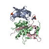
| ||||||||
|---|---|---|---|---|---|---|---|---|---|
| 1 | 
| ||||||||
| Unit cell |
| ||||||||
| Details | tetramer molecule with tetrahaedral symmetry |
-Components
-Dipeptydil-peptidase I ... , 3 types, 3 molecules ABC
| #1: Protein | Mass: 13500.163 Da / Num. of mol.: 1 Source method: isolated from a genetically manipulated source Source: (gene. exp.) Homo sapiens (human) / Cell (production host): INSECT CELL(Invitrogen) / Cell line (production host): high five / Production host:  Trichoplusia ni (cabbage looper) / References: UniProt: P53634, dipeptidyl-peptidase I Trichoplusia ni (cabbage looper) / References: UniProt: P53634, dipeptidyl-peptidase I |
|---|---|
| #2: Protein | Mass: 18491.871 Da / Num. of mol.: 1 Source method: isolated from a genetically manipulated source Source: (gene. exp.) Homo sapiens (human) / Cell (production host): INSECT CELL(Invitrogen) / Cell line (production host): high five / Production host: Trichoplusia ni (cabbage looper) / References: UniProt: P53634, dipeptidyl-peptidase I |
| #3: Protein | Mass: 7583.444 Da / Num. of mol.: 1 Source method: isolated from a genetically manipulated source Source: (gene. exp.) Homo sapiens (human) / Cell (production host): INSECT CELL(Invitrogen) / Cell line (production host): high five / Production host: Trichoplusia ni (cabbage looper) / References: UniProt: P53634, dipeptidyl-peptidase I |
-Sugars , 1 types, 1 molecules 
| #4: Sugar | ChemComp-NAG / N-Acetylglucosamine Type: D-saccharide, beta linking / Mass: 221.208 Da / Num. of mol.: 1 Type: D-saccharide, beta linking / Mass: 221.208 Da / Num. of mol.: 1Source method: isolated from a genetically manipulated source Formula: C8H15NO6 |
|---|
-Non-polymers , 3 types, 468 molecules 


| #5: Chemical | Sulfate Mass: 96.063 Da / Num. of mol.: 3 / Source method: obtained synthetically / Formula: SO4 Mass: 96.063 Da / Num. of mol.: 3 / Source method: obtained synthetically / Formula: SO4#6: Chemical | ChemComp-CL / | Chloride Mass: 35.453 Da / Num. of mol.: 1 / Source method: obtained synthetically / Formula: Cl Mass: 35.453 Da / Num. of mol.: 1 / Source method: obtained synthetically / Formula: Cl#7: Water | ChemComp-HOH / | WaterMass: 18.015 Da / Num. of mol.: 464 / Source method: isolated from a natural source / Formula: H2O |
|---|
-Experimental details
-Experiment
| Experiment | Method: X-RAY DIFFRACTION / Number of used crystals: 1 |
|---|
- Sample preparation
| Crystal | Density Matthews: 2.78 Å3/Da / Density % sol: 55.7 % | ||||||||||||||||||||||||||||||
|---|---|---|---|---|---|---|---|---|---|---|---|---|---|---|---|---|---|---|---|---|---|---|---|---|---|---|---|---|---|---|---|
| Crystal grow | Temperature: 293 K / Method: vapor diffusion, sitting drop / pH: 5.6 Details: ammonium sulphate, sodium citrate, potassium/sodium tartrate, pH 5.6, VAPOR DIFFUSION, SITTING DROP, temperature 293K | ||||||||||||||||||||||||||||||
| Crystal grow | *PLUS | ||||||||||||||||||||||||||||||
| Components of the solutions | *PLUS
|
-Data collection
| Diffraction | Mean temperature: 100 K |
|---|---|
| Diffraction source | Source: SYNCHROTRON / Site: ELETTRA  / Beamline: 5.2R / Wavelength: 1 Å / Beamline: 5.2R / Wavelength: 1 Å |
| Detector | Type: MARRESEARCH / Detector: AREA DETECTOR / Date: Jun 30, 1998 |
| Radiation | Protocol: SINGLE WAVELENGTH / Monochromatic (M) / Laue (L): M / Scattering type: x-ray |
| Radiation wavelength | Wavelength: 1 Å / Relative weight: 1 |
| Reflection | Resolution: 2.15→20 Å / Num. all: 23553 / Num. obs: 23553 / % possible obs: 97.6 % / Observed criterion σ(F): 2 / Observed criterion σ(I): 1 / Rmerge(I) obs: 0.07 |
| Reflection shell | Resolution: 2.15→2.18 Å / Num. unique all: 23353 / Rsym value: 0.249 / % possible all: 0.99 |
| Reflection | *PLUS Lowest resolution: 20 Å / Num. measured all: 96833 / Rmerge(I) obs: 0.07 |
| Reflection shell | *PLUS % possible obs: 99 % / Rmerge(I) obs: 0.249 |
- Processing
| Software |
| |||||||||||||||||||||||||
|---|---|---|---|---|---|---|---|---|---|---|---|---|---|---|---|---|---|---|---|---|---|---|---|---|---|---|
| Refinement | Method to determine structure: MAD, MIR, MOL. REPL together / Resolution: 2.15→10 Å / Isotropic thermal model: isotropic / σ(F): 1 / Stereochemistry target values: Engh & Huber
| |||||||||||||||||||||||||
| Displacement parameters | Biso mean: 23.8 Å2 | |||||||||||||||||||||||||
| Refinement step | Cycle: LAST / Resolution: 2.15→10 Å
| |||||||||||||||||||||||||
| Refine LS restraints |
| |||||||||||||||||||||||||
| Refinement | *PLUS Rfactor Rwork: 0.186 | |||||||||||||||||||||||||
| Solvent computation | *PLUS | |||||||||||||||||||||||||
| Displacement parameters | *PLUS |
