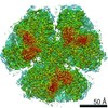
 Movie
Movie Controller


+ Open data

- Basic information
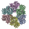
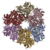
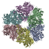
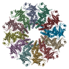
| Entry | Database: PDB / ID: 1k32 | ||||||
|---|---|---|---|---|---|---|---|
| Title | Crystal structure of the tricorn protease | ||||||
 Components Components | tricorn protease Proteasome endopeptidase complex Proteasome endopeptidase complex | ||||||
 Keywords Keywords | HYDROLASE / protein degradation / substrate gating / serine protease / beta propeller / proteasome | ||||||
| Function / homology |  Function and homology informationHydrolases; Acting on peptide bonds (peptidases); Serine endopeptidases / serine-type peptidase activity / proteolysis / cytoplasm Function and homology informationHydrolases; Acting on peptide bonds (peptidases); Serine endopeptidases / serine-type peptidase activity / proteolysis / cytoplasmSimilarity search - Function | ||||||
| Biological species |   Thermoplasma acidophilum (acidophilic) Thermoplasma acidophilum (acidophilic) | ||||||
| Method | X-RAY DIFFRACTION / SYNCHROTRON / MIR, MAD, NCS Averaging / Resolution: 2 Å | ||||||
 Authors Authors | Brandstetter, H. / Kim, J.-S. / Groll, M. / Huber, R. | ||||||
 Citation Citation | Journal: Nature / Year: 2001 Title: Crystal structure of the tricorn protease reveals a protein disassembly line. Authors: Brandstetter, H. / Kim, J.S. / Groll, M. / Huber, R. | ||||||
| History |
| ||||||
| Remark 600 | HETEROGEN HOH 2001-2399 ARE ASSOCIATED WITH CHAIN A. HOH 3001-3399 ARE ASSOCIATED WITH CHAIN B. HOH ...HETEROGEN HOH 2001-2399 ARE ASSOCIATED WITH CHAIN A. HOH 3001-3399 ARE ASSOCIATED WITH CHAIN B. HOH 4001-4399 ARE ASSOCIATED WITH CHAIN C. HOH 5001-5399 ARE ASSOCIATED WITH CHAIN D. HOH 6001-6399 ARE ASSOCIATED WITH CHAIN E. HOH 7001-7399 ARE ASSOCIATED WITH CHAIN F. |
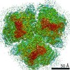
- Structure visualization
| Structure viewer | Molecule: MolmilJmol/JSmol |
|---|
- Downloads & links
-Download
| PDBx/mmCIF format | 1k32.cif.gz | 1.2 MB | Display | PDBx/mmCIF format |
|---|---|---|---|---|
| PDB format | pdb1k32.ent.gz | 1004.7 KB | Display | PDB format |
| PDBx/mmJSON format | 1k32.json.gz | Tree view | PDBx/mmJSON format | |
| Others |  Other downloads Other downloads |
-Validation report
| Arichive directory | https://data.pdbj.org/pub/pdb/validation_reports/k3/1k32ftp://data.pdbj.org/pub/pdb/validation_reports/k3/1k32 | HTTPS FTP |
|---|
-Related structure data
| Similar structure data |
|---|


-Links
 PDBj
PDBj
- Assembly
| Deposited unit | 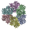
| ||||||||
|---|---|---|---|---|---|---|---|---|---|
| 1 |
| ||||||||
| Unit cell |
| ||||||||
| Details | homohexamer following 3-2 symmetry, completely contained in the coordinate file |
-Components
| #1: Protein | Proteasome endopeptidase complex Mass: 118680.055 Da / Num. of mol.: 6 Source method: isolated from a genetically manipulated source Source: (gene. exp.) Thermoplasma acidophilum (acidophilic) / Gene: TA1490 / Plasmid: pRSET-tric / Species (production host): Escherichia coli / Production host:  Escherichia coli BL21(DE3) (bacteria) / Strain (production host): BL21(DE3) Escherichia coli BL21(DE3) (bacteria) / Strain (production host): BL21(DE3)References: UniProt: P96086, Hydrolases; Acting on peptide bonds (peptidases); Serine endopeptidases#2: Water | ChemComp-HOH / | Water Mass: 18.015 Da / Num. of mol.: 2394 / Source method: isolated from a natural source / Formula: H2O Mass: 18.015 Da / Num. of mol.: 2394 / Source method: isolated from a natural source / Formula: H2O |
|---|
-Experimental details
-Experiment
| Experiment | Method: X-RAY DIFFRACTION / Number of used crystals: 1 |
|---|
- Sample preparation
| Crystal | Density Matthews: 2.54 Å3/Da / Density % sol: 51.56 % | ||||||||||||||||||
|---|---|---|---|---|---|---|---|---|---|---|---|---|---|---|---|---|---|---|---|
| Crystal grow | Temperature: 291 K / Method: vapor diffusion, hanging drop / pH: 6 Details: 25% isopropanol, 0.1 M MES, pH 6.0, VAPOR DIFFUSION, HANGING DROP, temperature 291K | ||||||||||||||||||
| Crystal | *PLUS Density % sol: 51 % | ||||||||||||||||||
| Crystal grow | *PLUS Temperature: 20 ℃ | ||||||||||||||||||
| Components of the solutions | *PLUS
|
-Data collection
| Diffraction | Mean temperature: 100 K |
|---|---|
| Diffraction source | Source: SYNCHROTRON / Site: ESRF  / Beamline: ID14-4 / Wavelength: 0.939 Å / Beamline: ID14-4 / Wavelength: 0.939 Å |
| Detector | Type: ADSC QUANTUM 4 / Detector: CCD / Date: Dec 10, 2000 |
| Radiation | Monochromator: Si 111 CHANNEL / Protocol: SINGLE WAVELENGTH / Monochromatic (M) / Laue (L): M / Scattering type: x-ray |
| Radiation wavelength | Wavelength: 0.939 Å / Relative weight: 1 |
| Reflection | Resolution: 2→20 Å / Num. all: 475881 / Num. obs: 394093 / % possible obs: 82.8 % / Observed criterion σ(F): 2 / Observed criterion σ(I): 3 / Redundancy: 2 % / Biso Wilson estimate: 26.2 Å2 / Rmerge(I) obs: 0.102 / Rsym value: 0.102 / Net I/σ(I): 6.7 |
| Reflection shell | Resolution: 2→2.05 Å / Redundancy: 1.2 % / Rmerge(I) obs: 0.313 / Mean I/σ(I) obs: 1.9 / Num. unique all: 394093 / Rsym value: 0.313 / % possible all: 68.1 |
| Reflection | *PLUS Rmerge(I) obs: 0.208 |
| Reflection shell | *PLUS Rmerge(I) obs: 0.313 |
- Processing
| Software |
| ||||||||||||||||||||||||||||||
|---|---|---|---|---|---|---|---|---|---|---|---|---|---|---|---|---|---|---|---|---|---|---|---|---|---|---|---|---|---|---|---|
| Refinement | Method to determine structure: MIR, MAD, NCS Averaging / Resolution: 2→20 Å Cross valid method: direct space free density correlation reciprocal free R σ(F): 0 / Stereochemistry target values: Engh & Huber
| ||||||||||||||||||||||||||||||
| Displacement parameters | Biso mean: 30.74 Å2 | ||||||||||||||||||||||||||||||
| Refinement step | Cycle: LAST / Resolution: 2→20 Å
| ||||||||||||||||||||||||||||||
| Refine LS restraints |
| ||||||||||||||||||||||||||||||
| LS refinement shell | Resolution: 2→2.04 Å
| ||||||||||||||||||||||||||||||
| Refinement | *PLUS Rfactor obs: 0.248 / Rfactor Rfree: 0.259 / Rfactor Rwork: 0.248 | ||||||||||||||||||||||||||||||
| Solvent computation | *PLUS | ||||||||||||||||||||||||||||||
| Displacement parameters | *PLUS | ||||||||||||||||||||||||||||||
| Refine LS restraints | *PLUS
| ||||||||||||||||||||||||||||||
| LS refinement shell | *PLUS Rfactor Rfree: 0.323 / Rfactor Rwork: 0.289 |
