 Movie
Movie Controller

[English] 日本語
 Yorodumi
- PDB-1k2x: Crystal structure of putative asparaginase encoded by Escherichia... -
+ Open data

- Basic information
| Entry | Database: PDB / ID: 1k2x | ||||||
|---|---|---|---|---|---|---|---|
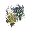
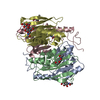
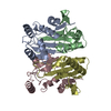
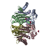
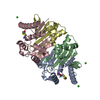
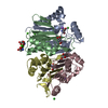
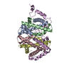
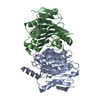
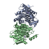
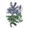
| Title | Crystal structure of putative asparaginase encoded by Escherichia coli ybiK gene | ||||||
 Components Components | (Putative L-asparaginase Asparaginase) x 2 Asparaginase) x 2 | ||||||
 Keywords Keywords | HYDROLASE / Ntn Hydrolase / Asparginase / Autoproteolysis | ||||||
| Function / homology |  Function and homology informationbeta-aspartyl-peptidase / asparaginase activity / beta-aspartyl-peptidase activity / protein autoprocessing / hydrolase activity / cytoplasm Function and homology informationbeta-aspartyl-peptidase / asparaginase activity / beta-aspartyl-peptidase activity / protein autoprocessing / hydrolase activity / cytoplasmSimilarity search - Function | ||||||
| Biological species |  Escherichia coli (E. coli) Escherichia coli (E. coli) | ||||||
| Method | X-RAY DIFFRACTION / SYNCHROTRON / MOLECULAR REPLACEMENT / Resolution: 1.65 Å | ||||||
 Authors Authors | Borek, D. / Jaskolski, M. | ||||||
 Citation Citation | Journal: Acta Crystallogr.,Sect.D / Year: 2008 Title: Crystal packing of plant-type L-asparaginase from Escherichia coli. Authors: Michalska, K. / Borek, D. / Hernandez-Santoyo, A. / Jaskolski, M. #1: Journal: Acta Crystallogr.,Sect.D / Year: 2000Title: Crystallization and Preliminary Crystallographic Studies of a New L-asparaginase Encoded by the Escherichia coli Genome Authors: Borek, D. / Jaskolski, M. #2: Journal: Nature / Year: 1995Title: A Protein Catalytic Framework with an N-terminal Nucleophile is Capable of Self-activation Authors: Brannigan, J.A. / Dodson, G. / Duggleby, H.J. / Moody, P.C. / Smith, J.L. / Tomchick, D.R. / Murzin, A.G. #3: Journal: Protein Sci. / Year: 1998Title: Crystal Structure of Glycosylasparaginase from Flavobacterium Meningosepticum Authors: Xuan, J. / Tarentino, A.L. / Grimwood, B.G. / Plummer Jr., T.H. / Cui, T. / Guan, C. / Van Roey, P. #4: Journal: Nat.Struct.Mol.Biol. / Year: 1995Title: Three-dimensional Structure of Human Lysosomal Aspartylglucosaminidase Authors: Oinonen, C. / Tikkanen, R. / Rouvinen, J. / Peltonen, L. #5: Journal: J.Biol.Chem. / Year: 1998Title: Crystal Structures of Flavobacterium Glycosylasparaginase. An N-terminal Nucleophile Hydrolase Activated by Intramolecular Proteolysis Authors: Guo, H.C. / Xu, Q. / Buckley, D. / Guan, C. #6: Journal: Cell(Cambridge,Mass.) / Year: 1999Title: Structural Insights into the Mechanism of Intramolecular Proteolysis Authors: Xu, Q. / Buckley, D. / Guan, C. / Guo, H.C. | ||||||
| History |
|
- Structure visualization
| Structure viewer | Molecule: MolmilJmol/JSmol |
|---|
- Downloads & links
-Download
| PDBx/mmCIF format | 1k2x.cif.gz | 136.4 KB | Display | PDBx/mmCIF format |
|---|---|---|---|---|
| PDB format | pdb1k2x.ent.gz | 104.6 KB | Display | PDB format |
| PDBx/mmJSON format | 1k2x.json.gz | Tree view | PDBx/mmJSON format | |
| Others |  Other downloads Other downloads |
-Validation report
| Arichive directory | https://data.pdbj.org/pub/pdb/validation_reports/k2/1k2xftp://data.pdbj.org/pub/pdb/validation_reports/k2/1k2x | HTTPS FTP |
|---|
-Related structure data
| Related structure data |  1jn9C  2zakC  2gawS C: citing same article ( S: Starting model for refinement |
|---|---|
| Similar structure data |


-Links
 PDBj
PDBj


- Assembly
| Deposited unit | 
| ||||||||
|---|---|---|---|---|---|---|---|---|---|
| 1 |
| ||||||||
| Unit cell |
| ||||||||
| Details | The biological assembly is a heterotetramer generated by non-crystallographical symmetry from two alpha+beta heterodimers. |
-Components
| #1: Protein | Asparaginase / E.C.3.5.1.1 / L-asparagine Amidohydrolase Mass: 18958.693 Da / Num. of mol.: 2 / Fragment: N-TERMINUS (RESIDUES 2-178) Source method: isolated from a genetically manipulated source Source: (gene. exp.) Escherichia coli (E. coli) / Gene: ybiK / Plasmid: pET11d / Production host: Escherichia coli (E. coli) / Strain (production host): JM109 / References: UniProt: P37595, asparaginase#2: Protein | Asparaginase / E.C.3.5.1.1 / L-asparagine AmidohydrolaseMass: 14428.146 Da / Num. of mol.: 2 / Fragment: C-TERMINUS (RESIDUES 179-321) Source method: isolated from a genetically manipulated source Source: (gene. exp.) Escherichia coli (E. coli) / Gene: ybiK / Plasmid: pET11d / Production host: Escherichia coli (E. coli) / Strain (production host): JM109 / References: UniProt: P37595, asparaginase#3: Chemical |   Mass: 22.990 Da / Num. of mol.: 2 / Source method: obtained synthetically / Formula: Na Mass: 22.990 Da / Num. of mol.: 2 / Source method: obtained synthetically / Formula: Na#4: Chemical | ChemComp-CL / Chloride  Mass: 35.453 Da / Num. of mol.: 9 / Source method: obtained synthetically / Formula: Cl Mass: 35.453 Da / Num. of mol.: 9 / Source method: obtained synthetically / Formula: Cl#5: Water | ChemComp-HOH / | Water Mass: 18.015 Da / Num. of mol.: 575 / Source method: isolated from a natural source / Formula: H2O Mass: 18.015 Da / Num. of mol.: 575 / Source method: isolated from a natural source / Formula: H2O |
|---|
-Experimental details
-Experiment
| Experiment | Method: X-RAY DIFFRACTION / Number of used crystals: 1 |
|---|
- Sample preparation
| Crystal | Density Matthews: 2.16 Å3/Da / Density % sol: 43.18 % | |||||||||||||||||||||||||||||||||||
|---|---|---|---|---|---|---|---|---|---|---|---|---|---|---|---|---|---|---|---|---|---|---|---|---|---|---|---|---|---|---|---|---|---|---|---|---|
| Crystal grow | Temperature: 292 K / Method: vapor diffusion, hanging drop / pH: 8.5 Details: PEG 4000, PEG 400, magnesium chloride, Tris-HCl pH 8.5, VAPOR DIFFUSION, HANGING DROP, temperature 292K | |||||||||||||||||||||||||||||||||||
| Crystal grow | *PLUS Method: vapor diffusion | |||||||||||||||||||||||||||||||||||
| Components of the solutions | *PLUS
|
-Data collection
| Diffraction | Mean temperature: 100 K |
|---|---|
| Diffraction source | Source: SYNCHROTRON / Site: EMBL/DESY, HAMBURG  / Beamline: X31 / Wavelength: 1.0442 Å / Beamline: X31 / Wavelength: 1.0442 Å |
| Detector | Type: MARRESEARCH / Detector: IMAGE PLATE / Date: Nov 4, 1999 / Details: Toroidal mirror |
| Radiation | Monochromator: Double-crystal monochromator / Protocol: SINGLE WAVELENGTH / Monochromatic (M) / Laue (L): M / Scattering type: x-ray |
| Radiation wavelength | Wavelength: 1.0442 Å / Relative weight: 1 |
| Reflection | Resolution: 1.65→20 Å / Num. all: 70249 / Num. obs: 70249 / Observed criterion σ(F): 0 / Observed criterion σ(I): -3 / Redundancy: 5.6 % / Rmerge(I) obs: 0.075 / Net I/σ(I): 16.8 |
| Reflection shell | Resolution: 1.65→1.68 Å / Redundancy: 5.5 % / Rmerge(I) obs: 0.476 / Mean I/σ(I) obs: 2.5 / % possible all: 89.9 |
- Processing
| Software |
| |||||||||||||||||||||||||||||||||||||||||||||||||||||||||||||||||||||||||||||||||||||||||||||||||||||||||||||||||||||||||||||||||||||||||||||||||||||||||||
|---|---|---|---|---|---|---|---|---|---|---|---|---|---|---|---|---|---|---|---|---|---|---|---|---|---|---|---|---|---|---|---|---|---|---|---|---|---|---|---|---|---|---|---|---|---|---|---|---|---|---|---|---|---|---|---|---|---|---|---|---|---|---|---|---|---|---|---|---|---|---|---|---|---|---|---|---|---|---|---|---|---|---|---|---|---|---|---|---|---|---|---|---|---|---|---|---|---|---|---|---|---|---|---|---|---|---|---|---|---|---|---|---|---|---|---|---|---|---|---|---|---|---|---|---|---|---|---|---|---|---|---|---|---|---|---|---|---|---|---|---|---|---|---|---|---|---|---|---|---|---|---|---|---|---|---|---|
| Refinement | Method to determine structure: MOLECULAR REPLACEMENT Starting model: PDB ENTRY 2GAW, polyalanine model Resolution: 1.65→19.5 Å / SU B: 2.286 / SU ML: 0.077 / TLS residual ADP flag: LIKELY RESIDUAL / Isotropic thermal model: Isotropic / Cross valid method: THROUGHOUT / ESU R: 0.085 / ESU R Free: 0.088 / Stereochemistry target values: MAXIMUM LIKELIHOOD / Details: HYDROGENS HAVE BEEN ADDED IN THE RIDING POSITIONS
| |||||||||||||||||||||||||||||||||||||||||||||||||||||||||||||||||||||||||||||||||||||||||||||||||||||||||||||||||||||||||||||||||||||||||||||||||||||||||||
| Solvent computation | Ion probe radii: 0.8 Å / Shrinkage radii: 0.8 Å / VDW probe radii: 1.4 Å / Solvent model: BABINET MODEL WITH MASK | |||||||||||||||||||||||||||||||||||||||||||||||||||||||||||||||||||||||||||||||||||||||||||||||||||||||||||||||||||||||||||||||||||||||||||||||||||||||||||
| Displacement parameters | Biso mean: 10.18 Å2
| |||||||||||||||||||||||||||||||||||||||||||||||||||||||||||||||||||||||||||||||||||||||||||||||||||||||||||||||||||||||||||||||||||||||||||||||||||||||||||
| Refine analyze |
| |||||||||||||||||||||||||||||||||||||||||||||||||||||||||||||||||||||||||||||||||||||||||||||||||||||||||||||||||||||||||||||||||||||||||||||||||||||||||||
| Refinement step | Cycle: LAST / Resolution: 1.65→19.5 Å
| |||||||||||||||||||||||||||||||||||||||||||||||||||||||||||||||||||||||||||||||||||||||||||||||||||||||||||||||||||||||||||||||||||||||||||||||||||||||||||
| Refine LS restraints |
| |||||||||||||||||||||||||||||||||||||||||||||||||||||||||||||||||||||||||||||||||||||||||||||||||||||||||||||||||||||||||||||||||||||||||||||||||||||||||||
| LS refinement shell | Resolution: 1.65→1.694 Å / Total num. of bins used: 20 /
| |||||||||||||||||||||||||||||||||||||||||||||||||||||||||||||||||||||||||||||||||||||||||||||||||||||||||||||||||||||||||||||||||||||||||||||||||||||||||||
| Refinement TLS params. | Method: refined / Refine-ID: X-RAY DIFFRACTION
| |||||||||||||||||||||||||||||||||||||||||||||||||||||||||||||||||||||||||||||||||||||||||||||||||||||||||||||||||||||||||||||||||||||||||||||||||||||||||||
| Refinement TLS group |
| |||||||||||||||||||||||||||||||||||||||||||||||||||||||||||||||||||||||||||||||||||||||||||||||||||||||||||||||||||||||||||||||||||||||||||||||||||||||||||
| Software | *PLUS Version: 5 / Classification: refinement | |||||||||||||||||||||||||||||||||||||||||||||||||||||||||||||||||||||||||||||||||||||||||||||||||||||||||||||||||||||||||||||||||||||||||||||||||||||||||||
| Refinement | *PLUS | |||||||||||||||||||||||||||||||||||||||||||||||||||||||||||||||||||||||||||||||||||||||||||||||||||||||||||||||||||||||||||||||||||||||||||||||||||||||||||
| Solvent computation | *PLUS | |||||||||||||||||||||||||||||||||||||||||||||||||||||||||||||||||||||||||||||||||||||||||||||||||||||||||||||||||||||||||||||||||||||||||||||||||||||||||||
| Displacement parameters | *PLUS |
