 Movie
Movie Controller


+ Open data

- Basic information
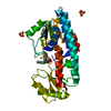
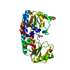
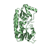
| Entry | Database: PDB / ID: 1k2v | ||||||
|---|---|---|---|---|---|---|---|
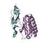
| Title | E. COLI PERIPLASMIC PROTEIN FHUD COMPLEXED WITH DESFERAL | ||||||
 Components Components | Ferrichrome-binding periplasmic protein | ||||||
 Keywords Keywords | METAL TRANSPORT / two domains / alpha helix linker | ||||||
| Function / homology |  Function and homology information Function and homology informationiron ion import across plasma membrane / ATP-binding cassette (ABC) transporter complex, substrate-binding subunit-containing / outer membrane-bounded periplasmic space Similarity search - Function | ||||||
| Biological species |   Escherichia coli (E. coli) Escherichia coli (E. coli) | ||||||
| Method | X-RAY DIFFRACTION / SYNCHROTRON / MOLECULAR REPLACEMENT / Resolution: 1.97 Å | ||||||
 Authors Authors | Clarke, T.E. / Braun, V. / Winkelmann, G. / Tari, L.W. / Vogel, H.J. | ||||||
 Citation Citation | Journal: J.Biol.Chem. / Year: 2002 Title: X-ray crystallographic structures of the Escherichia coli periplasmic protein FhuD bound to hydroxamate-type siderophores and the antibiotic albomycin. Authors: Clarke, T.E. / Braun, V. / Winkelmann, G. / Tari, L.W. / Vogel, H.J. | ||||||
| History |
|
- Structure visualization
| Structure viewer | Molecule: MolmilJmol/JSmol |
|---|
- Downloads & links
-Download
| PDBx/mmCIF format | 1k2v.cif.gz | 67.1 KB | Display | PDBx/mmCIF format |
|---|---|---|---|---|
| PDB format | pdb1k2v.ent.gz | 47.8 KB | Display | PDB format |
| PDBx/mmJSON format | 1k2v.json.gz | Tree view | PDBx/mmJSON format | |
| Others |  Other downloads Other downloads |
-Validation report
| Arichive directory | https://data.pdbj.org/pub/pdb/validation_reports/k2/1k2vftp://data.pdbj.org/pub/pdb/validation_reports/k2/1k2v | HTTPS FTP |
|---|
-Related structure data
| Related structure data |  1eszC 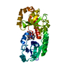 1k7sC 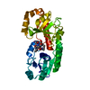 1efdS S: Starting model for refinement C: citing same article ( |
|---|---|
| Similar structure data |


-Links
 PDBj
PDBj- Assembly
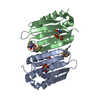
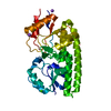
| Deposited unit | 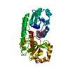
| ||||||||
|---|---|---|---|---|---|---|---|---|---|
| 1 |
| ||||||||
| Unit cell |
|
-Components
| #1: Protein | Mass: 29694.203 Da / Num. of mol.: 1 Source method: isolated from a genetically manipulated source Source: (gene. exp.) Escherichia coli (E. coli) / Gene: fhuD / Plasmid: pMR21 / Species (production host): Escherichia coli / Production host: Escherichia coli BL21(DE3) (bacteria) / Strain (production host): BL21(DE3) / References: UniProt: P07822 |
|---|---|
| #2: Chemical | ChemComp-DEF / 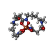  Mass: 721.622 Da / Num. of mol.: 1 / Source method: obtained synthetically / Formula: C27H49FeN6O11S Mass: 721.622 Da / Num. of mol.: 1 / Source method: obtained synthetically / Formula: C27H49FeN6O11S |
| #3: Water | ChemComp-HOH / Water Mass: 18.015 Da / Num. of mol.: 110 / Source method: isolated from a natural source / Formula: H2O Mass: 18.015 Da / Num. of mol.: 110 / Source method: isolated from a natural source / Formula: H2O |
-Experimental details
-Experiment
| Experiment | Method: X-RAY DIFFRACTION / Number of used crystals: 1 |
|---|
- Sample preparation
| Crystal | Density Matthews: 3.26 Å3/Da / Density % sol: 62.25 % | ||||||||||||||||||||||||||||||||||||
|---|---|---|---|---|---|---|---|---|---|---|---|---|---|---|---|---|---|---|---|---|---|---|---|---|---|---|---|---|---|---|---|---|---|---|---|---|---|
| Crystal grow | Temperature: 298 K / Method: vapor diffusion, hanging drop / pH: 4.6 Details: Na/K phosphate, HEPES, pH 4.6, VAPOR DIFFUSION, HANGING DROP, temperature 298K | ||||||||||||||||||||||||||||||||||||
| Crystal grow | *PLUS pH: 7.5 | ||||||||||||||||||||||||||||||||||||
| Components of the solutions | *PLUS
|
-Data collection
| Diffraction | Mean temperature: 100 K |
|---|---|
| Diffraction source | Source: SYNCHROTRON / Site: SSRL  / Beamline: BL7-1 / Beamline: BL7-1 |
| Detector | Type: ADSC QUANTUM 4 / Detector: CCD / Date: Dec 6, 2000 |
| Radiation | Protocol: SINGLE WAVELENGTH / Monochromatic (M) / Laue (L): M / Scattering type: x-ray |
| Radiation wavelength | Relative weight: 1 |
| Reflection | Resolution: 1.97→30 Å / Num. all: 25748 / Num. obs: 22886 / % possible obs: 88.9 % / Observed criterion σ(F): 1 / Observed criterion σ(I): 2 / Redundancy: 99.1 % / Rmerge(I) obs: 0.049 / Net I/σ(I): 21.8 |
| Reflection shell | Resolution: 1.97→2.04 Å / Redundancy: 97.8 % / Rmerge(I) obs: 0.205 / % possible all: 88.9 |
| Reflection | *PLUS Highest resolution: 2 Å / Lowest resolution: 30 Å / % possible obs: 99.1 % / Rmerge(I) obs: 0.049 |
| Reflection shell | *PLUS % possible obs: 97.8 % / Rmerge(I) obs: 0.205 / Mean I/σ(I) obs: 3.8 |
- Processing
| Software |
| |||||||||||||||||||||||||
|---|---|---|---|---|---|---|---|---|---|---|---|---|---|---|---|---|---|---|---|---|---|---|---|---|---|---|
| Refinement | Method to determine structure: MOLECULAR REPLACEMENT Starting model: PDB ENTRY 1EFD Resolution: 1.97→30 Å / Cross valid method: THROUGHOUT / σ(F): 1 / σ(I): 2 / Stereochemistry target values: MAXIMUM LIKELYHOOD FUNCTION
| |||||||||||||||||||||||||
| Refinement step | Cycle: LAST / Resolution: 1.97→30 Å
| |||||||||||||||||||||||||
| Refine LS restraints |
| |||||||||||||||||||||||||
| LS refinement shell | Resolution: 1.97→2.04 Å / Rfactor Rfree error: 0.021
| |||||||||||||||||||||||||
| Refinement | *PLUS Highest resolution: 2 Å / Lowest resolution: 30 Å / Rfactor obs: 0.22 / Rfactor Rfree: 0.242 / Rfactor Rwork: 0.22 | |||||||||||||||||||||||||
| Solvent computation | *PLUS | |||||||||||||||||||||||||
| Displacement parameters | *PLUS | |||||||||||||||||||||||||
| LS refinement shell | *PLUS Rfactor Rfree: 0.241 / Rfactor Rwork: 0.22 |
