 Movie
Movie Controller

[English] 日本語
 Yorodumi
- PDB-1k1b: Crystal structure of the ankyrin repeat domain of Bcl-3: a unique... -
+ Open data

- Basic information
| Entry | Database: PDB / ID: 1k1b | ||||||
|---|---|---|---|---|---|---|---|
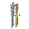
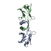
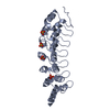
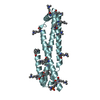
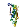
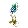
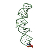
| Title | Crystal structure of the ankyrin repeat domain of Bcl-3: a unique member of the IkappaB protein family | ||||||
 Components Components | B-cell lymphoma 3-encoded protein | ||||||
 Keywords Keywords |  TRANSCRIPTION / Bcl-3 / NF-kappaB transcription factors / IkappaB proteins TRANSCRIPTION / Bcl-3 / NF-kappaB transcription factors / IkappaB proteins | ||||||
| Function / homology |  Function and homology information Function and homology informationBcl3-Bcl10 complex / follicular dendritic cell differentiation / Bcl3/NF-kappaB2 complex / regulation of DNA binding / marginal zone B cell differentiation / regulation of non-canonical NF-kappaB signal transduction / T cell apoptotic process / negative regulation of interleukin-8 production / germinal center formation / negative regulation of receptor signaling pathway via JAK-STAT ...Bcl3-Bcl10 complex / follicular dendritic cell differentiation / Bcl3/NF-kappaB2 complex / regulation of DNA binding / marginal zone B cell differentiation / regulation of non-canonical NF-kappaB signal transduction / T cell apoptotic process / negative regulation of interleukin-8 production / germinal center formation / negative regulation of receptor signaling pathway via JAK-STAT / humoral immune response mediated by circulating immunoglobulin / negative regulation of T cell apoptotic process / antimicrobial humoral response / T-helper 2 cell differentiation / response to UV-C / T-helper 1 type immune response / negative regulation of NF-kappaB transcription factor activity / defense response to protozoan / positive regulation of interleukin-10 production / negative regulation of tumor necrosis factor production / DNA damage response, signal transduction by p53 class mediator / intrinsic apoptotic signaling pathway in response to DNA damage by p53 class mediator / canonical NF-kappaB signal transduction / spleen development / extracellular matrix organization / positive regulation of translation / response to virus / protein import into nucleus / histone deacetylase binding / transcription corepressor activity / positive regulation of type II interferon production / protein-macromolecule adaptor activity / midbody / regulation of apoptotic process / DNA-binding transcription factor binding / transcription coactivator activity / defense response to bacterium / intracellular membrane-bounded organelle / negative regulation of DNA-templated transcription / DNA damage response / negative regulation of apoptotic process / perinuclear region of cytoplasm / positive regulation of DNA-templated transcription / positive regulation of transcription by RNA polymerase II / protein-containing complex / nucleoplasm / nucleus / plasma membrane / cytosol / cytoplasmSimilarity search - Function | ||||||
| Biological species |  Homo sapiens (human) Homo sapiens (human) | ||||||
| Method | X-RAY DIFFRACTION / SYNCHROTRON / MOLECULAR REPLACEMENT / Resolution: 1.9 Å | ||||||
 Authors Authors | Michel, F. / Soler-Lopez, M. / Petosa, C. / Cramer, P. / Siebenlist, U. / Mueller, C.W. | ||||||
 Citation Citation | Journal: EMBO J. / Year: 2001 Title: Crystal structure of the ankyrin repeat domain of Bcl-3: a unique member of the IkappaB protein family. Authors: Michel, F. / Soler-Lopez, M. / Petosa, C. / Cramer, P. / Siebenlist, U. / Muller, C.W. | ||||||
| History |
|
- Structure visualization
| Structure viewer | Molecule: MolmilJmol/JSmol |
|---|
- Downloads & links
-Download
| PDBx/mmCIF format | 1k1b.cif.gz | 59.7 KB | Display | PDBx/mmCIF format |
|---|---|---|---|---|
| PDB format | pdb1k1b.ent.gz | 42.5 KB | Display | PDB format |
| PDBx/mmJSON format | 1k1b.json.gz | Tree view | PDBx/mmJSON format | |
| Others |  Other downloads Other downloads |
-Validation report
| Arichive directory | https://data.pdbj.org/pub/pdb/validation_reports/k1/1k1bftp://data.pdbj.org/pub/pdb/validation_reports/k1/1k1b | HTTPS FTP |
|---|
-Related structure data
| Related structure data |  1k1aC  1iknS C: citing same article ( S: Starting model for refinement |
|---|---|
| Similar structure data |


-Links
 PDBj
PDBj

- Assembly
| Deposited unit | 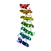
| ||||||||
|---|---|---|---|---|---|---|---|---|---|
| 1 |
| ||||||||
| Unit cell |
|
-Components
| #1: Protein | Mass: 25829.451 Da / Num. of mol.: 1 / Fragment: ANKYRIN REPEAT DOMAIN Source method: isolated from a genetically manipulated source Source: (gene. exp.) Homo sapiens (human) / Production host:  Escherichia coli (E. coli) / References: UniProt: P20749 Escherichia coli (E. coli) / References: UniProt: P20749 |
|---|---|
| #2: Water | ChemComp-HOH / Water Mass: 18.015 Da / Num. of mol.: 198 / Source method: isolated from a natural source / Formula: H2O Mass: 18.015 Da / Num. of mol.: 198 / Source method: isolated from a natural source / Formula: H2O |
-Experimental details
-Experiment
| Experiment | Method: X-RAY DIFFRACTION / Number of used crystals: 1 |
|---|
- Sample preparation
| Crystal | Density Matthews: 1.8 Å3/Da / Density % sol: 31.57 % | ||||||||||||||||||||||||||||||||||||||||||
|---|---|---|---|---|---|---|---|---|---|---|---|---|---|---|---|---|---|---|---|---|---|---|---|---|---|---|---|---|---|---|---|---|---|---|---|---|---|---|---|---|---|---|---|
| Crystal grow | Temperature: 293 K / Method: vapor diffusion, hanging drop / pH: 6 Details: PEG 6000, MES, pH 6.0, VAPOR DIFFUSION, HANGING DROP, temperature 293K | ||||||||||||||||||||||||||||||||||||||||||
| Crystal grow | *PLUS Temperature: 20 ℃ / pH: 5.8 | ||||||||||||||||||||||||||||||||||||||||||
| Components of the solutions | *PLUS
|
-Data collection
| Diffraction | Mean temperature: 100 K |
|---|---|
| Diffraction source | Source: SYNCHROTRON / Site: ESRF  / Beamline: ID14-1 / Wavelength: 0.934 Å / Beamline: ID14-1 / Wavelength: 0.934 Å |
| Detector | Type: MARRESEARCH / Detector: CCD / Date: Oct 28, 2000 |
| Radiation | Protocol: SINGLE WAVELENGTH / Monochromatic (M) / Laue (L): M / Scattering type: x-ray |
| Radiation wavelength | Wavelength: 0.934 Å / Relative weight: 1 |
| Reflection | Resolution: 1.9→40 Å / Num. all: 14381 / Num. obs: 14381 / % possible obs: 98.6 % / Observed criterion σ(F): 0 / Observed criterion σ(I): 0 / Redundancy: 3.41 % / Rsym value: 0.097 / Net I/σ(I): 15.1 |
| Reflection shell | Resolution: 1.9→1.97 Å / Redundancy: 2.68 % / Mean I/σ(I) obs: 3.15 / Num. unique all: 1378 / Rsym value: 0.26 / % possible all: 94.4 |
| Reflection | *PLUS Lowest resolution: 40 Å / Num. measured all: 49074 / Rmerge(I) obs: 0.097 |
| Reflection shell | *PLUS % possible obs: 94.4 % / Rmerge(I) obs: 0.26 |
- Processing
| Software |
| ||||||||||||||||||||||||||||||||
|---|---|---|---|---|---|---|---|---|---|---|---|---|---|---|---|---|---|---|---|---|---|---|---|---|---|---|---|---|---|---|---|---|---|
| Refinement | Method to determine structure: MOLECULAR REPLACEMENT Starting model: 1ikn Resolution: 1.9→20 Å / SU B: 3.61543 / SU ML: 0.10851 / Cross valid method: THROUGHOUT / σ(F): 0 / ESU R: 0.19561 / ESU R Free: 0.15851 / Stereochemistry target values: Engh & Huber Details: RESIDUES LEU182 AND SER307 PRESENT DOUBLE CONFORMATIONS.
| ||||||||||||||||||||||||||||||||
| Displacement parameters | Biso mean: 19.188 Å2
| ||||||||||||||||||||||||||||||||
| Refinement step | Cycle: LAST / Resolution: 1.9→20 Å
| ||||||||||||||||||||||||||||||||
| Refine LS restraints |
| ||||||||||||||||||||||||||||||||
| Software | *PLUS Name: REFMAC / Classification: refinement | ||||||||||||||||||||||||||||||||
| Refinement | *PLUS Highest resolution: 1.9 Å / Lowest resolution: 20 Å / σ(F): 0 / % reflection Rfree: 5 % | ||||||||||||||||||||||||||||||||
| Solvent computation | *PLUS | ||||||||||||||||||||||||||||||||
| Displacement parameters | *PLUS | ||||||||||||||||||||||||||||||||
| Refine LS restraints | *PLUS
|
