 Movie
Movie Controller

[English] 日本語
 Yorodumi
- PDB-1jwv: Crystal structure of G238A mutant of TEM-1 beta-lactamase in comp... -
+ Open data

- Basic information
| Entry | Database: PDB / ID: 1jwv | ||||||
|---|---|---|---|---|---|---|---|
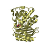
| Title | Crystal structure of G238A mutant of TEM-1 beta-lactamase in complex with a boronic acid inhibitor (sefb4) | ||||||
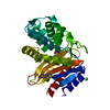
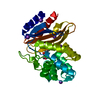
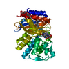
 Components Components | BETA-LACTAMASE TEM | ||||||
 Keywords Keywords |  HYDROLASE / TEM-1 / beta-lactamase / serine hydrolase HYDROLASE / TEM-1 / beta-lactamase / serine hydrolase | ||||||
| Function / homology |  Function and homology information Function and homology informationbeta-lactam antibiotic catabolic process / beta-lactamase activity / beta-lactamase / response to antibioticSimilarity search - Function | ||||||
| Biological species |  Escherichia coli (E. coli) Escherichia coli (E. coli) | ||||||
| Method | X-RAY DIFFRACTION / SYNCHROTRON / MOLECULAR REPLACEMENT / Resolution: 1.85 Å | ||||||
 Authors Authors | Wang, X. / Minasov, G. / Shoichet, B.K. | ||||||
 Citation Citation | Journal: J.Mol.Biol. / Year: 2002 Title: Evolution of an antibiotic resistance enzyme constrained by stability and activity trade-offs. Authors: Wang, X. / Minasov, G. / Shoichet, B.K. | ||||||
| History |
|
- Structure visualization
| Structure viewer | Molecule: MolmilJmol/JSmol |
|---|
- Downloads & links
-Download
| PDBx/mmCIF format | 1jwv.cif.gz | 78.5 KB | Display | PDBx/mmCIF format |
|---|---|---|---|---|
| PDB format | pdb1jwv.ent.gz | 55.5 KB | Display | PDB format |
| PDBx/mmJSON format | 1jwv.json.gz | Tree view | PDBx/mmJSON format | |
| Others |  Other downloads Other downloads |
-Validation report
| Arichive directory | https://data.pdbj.org/pub/pdb/validation_reports/jw/1jwvftp://data.pdbj.org/pub/pdb/validation_reports/jw/1jwv | HTTPS FTP |
|---|
-Related structure data
| Related structure data |  1jwpC 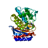 1jwzC  1btlS S: Starting model for refinement C: citing same article ( |
|---|---|
| Similar structure data |


-Links
 PDBj
PDBj


- Assembly
| Deposited unit | 
| ||||||||
|---|---|---|---|---|---|---|---|---|---|
| 1 |
| ||||||||
| Unit cell |
|
-Components
| #1: Protein | Mass: 28956.021 Da / Num. of mol.: 1 / Fragment: TEM-1 / Mutation: G238A Source method: isolated from a genetically manipulated source Source: (gene. exp.) Escherichia coli (E. coli) / Gene: bla / Plasmid: pAiter EX II / Production host: Escherichia coli (E. coli) / Strain (production host): SF120 / References: UniProt: P62593, beta-lactamase | ||||
|---|---|---|---|---|---|
| #2: Chemical | ChemComp-K /   Mass: 39.098 Da / Num. of mol.: 5 / Source method: obtained synthetically / Formula: K Mass: 39.098 Da / Num. of mol.: 5 / Source method: obtained synthetically / Formula: K#3: Chemical | ChemComp-CB4 / | 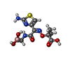  Mass: 330.125 Da / Num. of mol.: 1 / Source method: obtained synthetically / Formula: C10H15BN4O6S Mass: 330.125 Da / Num. of mol.: 1 / Source method: obtained synthetically / Formula: C10H15BN4O6S#4: Water | ChemComp-HOH / | Water Mass: 18.015 Da / Num. of mol.: 417 / Source method: isolated from a natural source / Formula: H2O Mass: 18.015 Da / Num. of mol.: 417 / Source method: isolated from a natural source / Formula: H2O |
-Experimental details
-Experiment
| Experiment | Method: X-RAY DIFFRACTION / Number of used crystals: 1 |
|---|
- Sample preparation
| Crystal | Density Matthews: 1.97 Å3/Da / Density % sol: 37.5 % | ||||||||||||||||||
|---|---|---|---|---|---|---|---|---|---|---|---|---|---|---|---|---|---|---|---|
| Crystal grow | Temperature: 295 K / Method: vapor diffusion, hanging drop / pH: 8 Details: sodium-potassium phosphate buffer, pH 8.0, VAPOR DIFFUSION, HANGING DROP, temperature 295.0K | ||||||||||||||||||
| Crystal grow | *PLUS Method: unknown | ||||||||||||||||||
| Components of the solutions | *PLUS
|
-Data collection
| Diffraction | Mean temperature: 100 K |
|---|---|
| Diffraction source | Source: SYNCHROTRON / Site: APS  / Beamline: 5ID-B / Wavelength: 1 Å / Beamline: 5ID-B / Wavelength: 1 Å |
| Detector | Type: MARRESEARCH / Detector: CCD / Date: Jun 6, 2001 / Details: mirrors |
| Radiation | Monochromator: graphite / Protocol: SINGLE WAVELENGTH / Monochromatic (M) / Laue (L): M / Scattering type: x-ray |
| Radiation wavelength | Wavelength: 1 Å / Relative weight: 1 |
| Reflection | Resolution: 1.85→20 Å / Num. all: 20097 / Num. obs: 20097 / % possible obs: 99.9 % / Observed criterion σ(F): 0 / Observed criterion σ(I): -3 / Redundancy: 6 % / Biso Wilson estimate: 22.2 Å2 / Rmerge(I) obs: 0.064 / Net I/σ(I): 25.99 |
| Reflection shell | Resolution: 1.85→1.92 Å / Redundancy: 5.9 % / Rmerge(I) obs: 0.121 / Mean I/σ(I) obs: 14.2 / Num. unique all: 1959 / % possible all: 99.6 |
| Reflection | *PLUS |
| Reflection shell | *PLUS % possible obs: 99.6 % / Num. unique obs: 1959 |
- Processing
| Software |
| |||||||||||||||||||||||||
|---|---|---|---|---|---|---|---|---|---|---|---|---|---|---|---|---|---|---|---|---|---|---|---|---|---|---|
| Refinement | Method to determine structure: MOLECULAR REPLACEMENT Starting model: 1BTL Resolution: 1.85→19 Å / Isotropic thermal model: Isotropic / Cross valid method: THROUGHOUT / σ(F): 0 / σ(I): 0 / Stereochemistry target values: Engh & Huber Details: Crystallographic conjugate gradient minimization refinement using maximum likelihood target for amplitudes
| |||||||||||||||||||||||||
| Displacement parameters | Biso mean: 14.3 Å2
| |||||||||||||||||||||||||
| Refine analyze |
| |||||||||||||||||||||||||
| Refinement step | Cycle: LAST / Resolution: 1.85→19 Å
| |||||||||||||||||||||||||
| Refine LS restraints |
| |||||||||||||||||||||||||
| LS refinement shell | Resolution: 1.85→1.92 Å / Total num. of bins used: 10
| |||||||||||||||||||||||||
| Refinement | *PLUS Lowest resolution: 20 Å / % reflection Rfree: 10 % / Rfactor obs: 0.165 | |||||||||||||||||||||||||
| Solvent computation | *PLUS | |||||||||||||||||||||||||
| Displacement parameters | *PLUS | |||||||||||||||||||||||||
| Refine LS restraints | *PLUS
| |||||||||||||||||||||||||
| LS refinement shell | *PLUS Rfactor obs: 0.224 |
