 Movie
Movie Controller


+ Open data

- Basic information
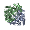
| Entry | Database: PDB / ID: 1jfd | ||||||
|---|---|---|---|---|---|---|---|
| Title | STRUCTURE OF INORGANIC PYROPHOSPHATASE | ||||||
 Components Components | INORGANIC PYROPHOSPHATASE | ||||||
 Keywords Keywords | HYDROLASE / ACID ANHYDRIDE HYDROLASE | ||||||
| Function / homology |  Function and homology information Function and homology informationinorganic triphosphate phosphatase activity / inorganic diphosphatase / inorganic diphosphate phosphatase activity / phosphate-containing compound metabolic process / magnesium ion binding / zinc ion binding / membrane / cytosolSimilarity search - Function | ||||||
| Biological species |  Escherichia coli (E. coli) Escherichia coli (E. coli) | ||||||
| Method | X-RAY DIFFRACTION / Resolution: 2.2 Å | ||||||
 Authors Authors | Oganesyan, V. / Avaeva, S.M. / Huber, R. / Harutyunyan, E.H. | ||||||
 Citation Citation | Journal: FEBS Lett. / Year: 1997 Title: Crystal structure of Escherichia coli inorganic pyrophosphatase complexed with SO4(2-). Ligand-induced molecular asymmetry. Authors: Avaeva, S. / Kurilova, S. / Nazarova, T. / Rodina, E. / Vorobyeva, N. / Sklyankina, V. / Grigorjeva, O. / Harutyunyan, E. / Oganessyan, V. / Wilson, K. / Dauter, Z. / Huber, R. / Mather, T. #1: Journal: FEBS Lett. / Year: 1994Title: X-Ray Crystallographic Studies of Recombinant Inorganic Pyrophosphatase from Escherichia Coli Authors: Oganessyan, V.Yu. / Kurilova, S.A. / Vorobyeva, N.N. / Nazarova, T.I. / Popov, A.N. / Lebedev, A.A. / Avaeva, S.M. / Harutyunyan, E.H. | ||||||
| History |
|
- Structure visualization
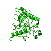
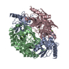
| Structure viewer | Molecule: MolmilJmol/JSmol |
|---|
- Downloads & links
-Download
| PDBx/mmCIF format | 1jfd.cif.gz | 82.8 KB | Display | PDBx/mmCIF format |
|---|---|---|---|---|
| PDB format | pdb1jfd.ent.gz | 63.8 KB | Display | PDB format |
| PDBx/mmJSON format | 1jfd.json.gz | Tree view | PDBx/mmJSON format | |
| Others |  Other downloads Other downloads |
-Validation report
| Arichive directory | https://data.pdbj.org/pub/pdb/validation_reports/jf/1jfdftp://data.pdbj.org/pub/pdb/validation_reports/jf/1jfd | HTTPS FTP |
|---|
-Related structure data
| Similar structure data |
|---|


-Links
 PDBj
PDBj- Assembly
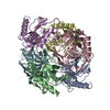
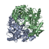
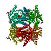
| Deposited unit | 
| ||||||||
|---|---|---|---|---|---|---|---|---|---|
| 1 | 
| ||||||||
| Unit cell |
| ||||||||
| Noncrystallographic symmetry (NCS) | NCS oper: (Code: given Matrix: (0.999803, 0.019396, -0.004307), Vector : |
-Components
| #1: Protein | / PYROPHOSPHATE HYDROLASE Mass: 19597.334 Da / Num. of mol.: 2 Source method: isolated from a genetically manipulated source Source: (gene. exp.) Escherichia coli (E. coli) / Strain: JM109 / Plasmid: PUC19Gene (production host): PYROPHOSPHATASE FROM ESCHERICHIA COLI Production host: Escherichia coli (E. coli) / References: UniProt: P0A7A9, inorganic diphosphatase#2: Chemical | Sulfate  Mass: 96.063 Da / Num. of mol.: 2 / Source method: obtained synthetically / Formula: SO4 Mass: 96.063 Da / Num. of mol.: 2 / Source method: obtained synthetically / Formula: SO4#3: Water | ChemComp-HOH / | Water Mass: 18.015 Da / Num. of mol.: 207 / Source method: isolated from a natural source / Formula: H2O Mass: 18.015 Da / Num. of mol.: 207 / Source method: isolated from a natural source / Formula: H2O |
|---|
-Experimental details
-Experiment
| Experiment | Method: X-RAY DIFFRACTION |
|---|
- Sample preparation
| Crystal | Density Matthews: 2.31 Å3/Da / Density % sol: 40 % | ||||||||||||||||||||||||||||||
|---|---|---|---|---|---|---|---|---|---|---|---|---|---|---|---|---|---|---|---|---|---|---|---|---|---|---|---|---|---|---|---|
| Crystal grow | *PLUS pH: 7.5 / Method: vapor diffusion, hanging drop | ||||||||||||||||||||||||||||||
| Components of the solutions | *PLUS
|
-Data collection
| Diffraction source | Wavelength: 1.09 |
|---|---|
| Detector | Type: MARRESEARCH / Detector: IMAGE PLATE AREA DETECTOR / Date: Apr 1, 1991 |
| Radiation | Monochromatic (M) / Laue (L): M / Scattering type: x-ray |
| Radiation wavelength | Wavelength: 1.09 Å / Relative weight: 1 |
| Reflection | Num. obs: 18662 / % possible obs: 99.2 % / Observed criterion σ(I): 3 / Rmerge(I) obs: 0.077 |
| Reflection | *PLUS Highest resolution: 2.196 Å / Num. measured all: 153537 |
- Processing
| Software |
| ||||||||||||||||||||||||||||||||||||||||||||||||||||||||||||||||||||||||||||||||||||
|---|---|---|---|---|---|---|---|---|---|---|---|---|---|---|---|---|---|---|---|---|---|---|---|---|---|---|---|---|---|---|---|---|---|---|---|---|---|---|---|---|---|---|---|---|---|---|---|---|---|---|---|---|---|---|---|---|---|---|---|---|---|---|---|---|---|---|---|---|---|---|---|---|---|---|---|---|---|---|---|---|---|---|---|---|---|
| Refinement | Resolution: 2.2→15 Å / σ(F): 3 Details: ESTIMATED COORD. ERROR 0.35 ANGSTROMS FINAL RMS COORD. SHIFT 0.002 ANGSTROMS
| ||||||||||||||||||||||||||||||||||||||||||||||||||||||||||||||||||||||||||||||||||||
| Displacement parameters | Biso mean: 21.34 Å2 | ||||||||||||||||||||||||||||||||||||||||||||||||||||||||||||||||||||||||||||||||||||
| Refine analyze | Luzzati coordinate error obs: 0.25 Å | ||||||||||||||||||||||||||||||||||||||||||||||||||||||||||||||||||||||||||||||||||||
| Refinement step | Cycle: LAST / Resolution: 2.2→15 Å
| ||||||||||||||||||||||||||||||||||||||||||||||||||||||||||||||||||||||||||||||||||||
| Refine LS restraints |
| ||||||||||||||||||||||||||||||||||||||||||||||||||||||||||||||||||||||||||||||||||||
| Software | *PLUS Name: REFMAC / Classification: refinement | ||||||||||||||||||||||||||||||||||||||||||||||||||||||||||||||||||||||||||||||||||||
| Refinement | *PLUS Rfactor obs: 0.192 | ||||||||||||||||||||||||||||||||||||||||||||||||||||||||||||||||||||||||||||||||||||
| Solvent computation | *PLUS | ||||||||||||||||||||||||||||||||||||||||||||||||||||||||||||||||||||||||||||||||||||
| Displacement parameters | *PLUS |
