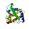
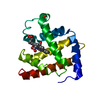
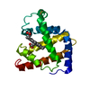
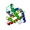
 Movie
Movie Controller


+ Open data

- Basic information
| Entry | Database: PDB / ID: 1jdo | ||||||
|---|---|---|---|---|---|---|---|
| Title | SPERM WHALE MYOGLOBIN (FERROUS, NITRIC OXIDE BOUND) | ||||||
 Components Components | MYOGLOBIN | ||||||
 Keywords Keywords | OXYGEN TRANSPORT / GLOBIN / HEME / OXYGEN STORAGE / NITRIC OXIDE | ||||||
| Function / homology |  Function and homology information Function and homology informationhydrogen peroxide mediated signaling pathway / oxygen carrier activity / oxygen binding / heme binding / metal ion bindingSimilarity search - Function | ||||||
| Biological species |  Physeter catodon (sperm whale) Physeter catodon (sperm whale) | ||||||
| Method | X-RAY DIFFRACTION / DIFFERENCE FOURIER / Resolution: 1.9 Å | ||||||
 Authors Authors | Brucker, E.A. / Phillips Jr., G.N. | ||||||
 Citation Citation | Journal: Proteins / Year: 1998 Title: Nitric oxide myoglobin: crystal structure and analysis of ligand geometry. Authors: Brucker, E.A. / Olson, J.S. / Ikeda-Saito, M. / Phillips Jr., G.N. | ||||||
| History |
|
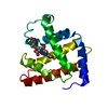
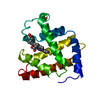
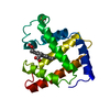
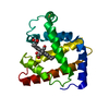
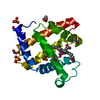
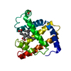
- Structure visualization
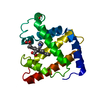
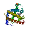
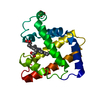
| Structure viewer | Molecule: MolmilJmol/JSmol |
|---|
- Downloads & links
-Download
| PDBx/mmCIF format | 1jdo.cif.gz | 44.6 KB | Display | PDBx/mmCIF format |
|---|---|---|---|---|
| PDB format | pdb1jdo.ent.gz | 33.7 KB | Display | PDB format |
| PDBx/mmJSON format | 1jdo.json.gz | Tree view | PDBx/mmJSON format | |
| Others |  Other downloads Other downloads |
-Validation report
| Arichive directory | https://data.pdbj.org/pub/pdb/validation_reports/jd/1jdoftp://data.pdbj.org/pub/pdb/validation_reports/jd/1jdo | HTTPS FTP |
|---|
-Related structure data
| Related structure data | 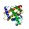 1hjtC 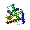 2splS S: Starting model for refinement C: citing same article ( |
|---|---|
| Similar structure data |


-Links
 PDBj
PDBj


- Assembly
| Deposited unit | 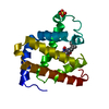
| ||||||||
|---|---|---|---|---|---|---|---|---|---|
| 1 |
| ||||||||
| Unit cell |
|
-Components
| #1: Protein | Mass: 17399.180 Da / Num. of mol.: 1 / Mutation: INS(M0), L29F, D122N Source method: isolated from a genetically manipulated source Source: (gene. exp.) Physeter catodon (sperm whale) / Production host:  Escherichia coli (E. coli) / References: UniProt: P02185 Escherichia coli (E. coli) / References: UniProt: P02185 |
|---|---|
| #2: Chemical | ChemComp-SO4 / Sulfate  Mass: 96.063 Da / Num. of mol.: 1 / Source method: obtained synthetically / Formula: SO4 Mass: 96.063 Da / Num. of mol.: 1 / Source method: obtained synthetically / Formula: SO4 |
| #3: Chemical | ChemComp-HEM / Heme B  Mass: 616.487 Da / Num. of mol.: 1 / Source method: obtained synthetically / Formula: C34H32FeN4O4 Mass: 616.487 Da / Num. of mol.: 1 / Source method: obtained synthetically / Formula: C34H32FeN4O4 |
| #4: Chemical | ChemComp-NO / Nitric oxide  Mass: 30.006 Da / Num. of mol.: 1 / Source method: obtained synthetically / Formula: NO Mass: 30.006 Da / Num. of mol.: 1 / Source method: obtained synthetically / Formula: NO |
| #5: Water | ChemComp-HOH / Water Mass: 18.015 Da / Num. of mol.: 138 / Source method: isolated from a natural source / Formula: H2O Mass: 18.015 Da / Num. of mol.: 138 / Source method: isolated from a natural source / Formula: H2O |
| Sequence details | THIS STRUCTURE HAS THREE DIFFERENCES RELATIVE TO THE NATIVE SPERM WHALE PROTEIN: AN INITIATOR ...THIS STRUCTURE HAS THREE DIFFERENCE |
-Experimental details
-Experiment
| Experiment | Method: X-RAY DIFFRACTION / Number of used crystals: 1 |
|---|
- Sample preparation
| Crystal | Density Matthews: 3.18 Å3/Da / Density % sol: 61.37 % |
|---|---|
| Crystal grow | pH: 9 / Details: pH 9. |
-Data collection
| Diffraction | Mean temperature: 295 K |
|---|---|
| Diffraction source | Source: ROTATING ANODE / Type: SIEMENS / Wavelength: 1.5418 |
| Detector | Type: RIGAKU / Detector: IMAGE PLATE / Date: Feb 14, 1997 |
| Radiation | Monochromatic (M) / Laue (L): M / Scattering type: x-ray |
| Radiation wavelength | Wavelength: 1.5418 Å / Relative weight: 1 |
| Reflection | Resolution: 1.9→30 Å / Num. obs: 17289 / % possible obs: 99 % / Redundancy: 4.2 % / Rmerge(I) obs: 0.064 / Net I/σ(I): 26.4 |
| Reflection shell | Resolution: 1.9→2 Å / Redundancy: 3.9 % / Rmerge(I) obs: 0.292 / Mean I/σ(I) obs: 5.5 / % possible all: 99.8 |
- Processing
| Software |
| |||||||||||||||||||||||||||||||||
|---|---|---|---|---|---|---|---|---|---|---|---|---|---|---|---|---|---|---|---|---|---|---|---|---|---|---|---|---|---|---|---|---|---|---|
| Refinement | Method to determine structure: DIFFERENCE FOURIER Starting model: PDB ENTRY 2SPL Resolution: 1.9→5 Å / Num. parameters: 5667 / Num. restraintsaints: 5401 / Stereochemistry target values: ENGH AND HUBER
| |||||||||||||||||||||||||||||||||
| Solvent computation | Solvent model: MOEWS & KRETSINGER, J.MOL.BIOL.91(1973)201-228 | |||||||||||||||||||||||||||||||||
| Refine analyze | Num. disordered residues: 0 / Occupancy sum hydrogen: 0 / Occupancy sum non hydrogen: 1382.3 | |||||||||||||||||||||||||||||||||
| Refinement step | Cycle: LAST / Resolution: 1.9→5 Å
| |||||||||||||||||||||||||||||||||
| Refine LS restraints |
|
