 Movie
Movie Controller


+ Open data

- Basic information
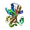
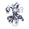
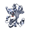
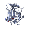
| Entry | Database: PDB / ID: 1jd3 | ||||||
|---|---|---|---|---|---|---|---|
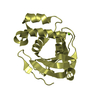
| Title | Chorismate lyase G90A mutant with bound product | ||||||
 Components Components | chorismate lyase | ||||||
 Keywords Keywords | LYASE / active site mutant / product complex | ||||||
| Function / homology |  Function and homology informationchorismate lyase / chorismate lyase activity / pyruvate biosynthetic process / ubiquinone biosynthetic process / cytosol Function and homology informationchorismate lyase / chorismate lyase activity / pyruvate biosynthetic process / ubiquinone biosynthetic process / cytosolSimilarity search - Function | ||||||
| Biological species |  Escherichia coli (E. coli) Escherichia coli (E. coli) | ||||||
| Method | X-RAY DIFFRACTION / FOURIER SYNTHESIS / Resolution: 2.03 Å | ||||||
 Authors Authors | Mayhew, M. / Smith, N. / Holden, M.J. / Gallagher, D.T. | ||||||
 Citation Citation | Journal: Arch.Biochem.Biophys. / Year: 2006 Title: Structural analysis of ligand binding and catalysis in chorismate lyase. Authors: Smith, N. / Roitberg, A.E. / Rivera, E. / Howard, A. / Holden, M.J. / Mayhew, M. / Kaistha, S. / Gallagher, D.T. | ||||||
| History |
|
- Structure visualization
| Structure viewer | Molecule: MolmilJmol/JSmol |
|---|
- Downloads & links
-Download
| PDBx/mmCIF format | 1jd3.cif.gz | 47.7 KB | Display | PDBx/mmCIF format |
|---|---|---|---|---|
| PDB format | pdb1jd3.ent.gz | 32.9 KB | Display | PDB format |
| PDBx/mmJSON format | 1jd3.json.gz | Tree view | PDBx/mmJSON format | |
| Others |  Other downloads Other downloads |
-Validation report
| Arichive directory | https://data.pdbj.org/pub/pdb/validation_reports/jd/1jd3ftp://data.pdbj.org/pub/pdb/validation_reports/jd/1jd3 | HTTPS FTP |
|---|
-Related structure data
| Related structure data | 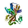 1tt8C 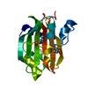 1xlrC  2ahcC 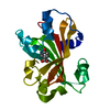 1g81S S: Starting model for refinement C: citing same article ( |
|---|---|
| Similar structure data |


-Links
 PDBj
PDBj- Assembly
| Deposited unit | 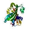
| ||||||||
|---|---|---|---|---|---|---|---|---|---|
| 1 |
| ||||||||
| Unit cell |
|
-Components
| #1: Protein | Mass: 18649.582 Da / Num. of mol.: 1 / Mutation: C14S,C81S,G90A Source method: isolated from a genetically manipulated source Source: (gene. exp.) Escherichia coli (E. coli) / Production host: Escherichia coli (E. coli) / References: UniProt: P26602, Lyases |
|---|---|
| #2: Chemical | ChemComp-PHB / 4-Hydroxybenzoic acid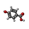  Mass: 138.121 Da / Num. of mol.: 1 / Source method: obtained synthetically / Formula: C7H6O3 Mass: 138.121 Da / Num. of mol.: 1 / Source method: obtained synthetically / Formula: C7H6O3 |
| #3: Water | ChemComp-HOH / Water Mass: 18.015 Da / Num. of mol.: 65 / Source method: isolated from a natural source / Formula: H2O Mass: 18.015 Da / Num. of mol.: 65 / Source method: isolated from a natural source / Formula: H2O |
-Experimental details
-Experiment
| Experiment | Method: X-RAY DIFFRACTION / Number of used crystals: 1 |
|---|
- Sample preparation
| Crystal | Density Matthews: 2.03 Å3/Da / Density % sol: 39.51 % |
|---|---|
| Crystal grow | Temperature: 297 K / Method: vapor diffusion, hanging drop / pH: 7 Details: PEG 4K, hepes, isopropanol, pH 7.0, VAPOR DIFFUSION, HANGING DROP, temperature 297K |
-Data collection
| Diffraction | Mean temperature: 298 K |
|---|---|
| Diffraction source | Source: ROTATING ANODE / Type: SIEMENS / Wavelength: 1.54 Å |
| Detector | Type: MARRESEARCH / Detector: IMAGE PLATE / Date: Mar 21, 2001 / Details: mirrors |
| Radiation | Protocol: SINGLE WAVELENGTH / Monochromatic (M) / Laue (L): M / Scattering type: x-ray |
| Radiation wavelength | Wavelength: 1.54 Å / Relative weight: 1 |
| Reflection | Resolution: 2→20 Å / Num. all: 9861 / Num. obs: 9861 / % possible obs: 94 % / Observed criterion σ(F): 0 / Observed criterion σ(I): 0 / Redundancy: 3.4 % / Biso Wilson estimate: 22.2 Å2 / Rmerge(I) obs: 0.06 / Net I/σ(I): 10.1 |
| Reflection shell | Resolution: 2→2.15 Å / Redundancy: 2.1 % / Rmerge(I) obs: 0.22 / % possible all: 83 |
- Processing
| Software |
| ||||||||||||||||||||||||||||||||||||
|---|---|---|---|---|---|---|---|---|---|---|---|---|---|---|---|---|---|---|---|---|---|---|---|---|---|---|---|---|---|---|---|---|---|---|---|---|---|
| Refinement | Method to determine structure: FOURIER SYNTHESIS Starting model: 1g81.pdb Resolution: 2.03→8 Å / Rfactor Rfree error: 0.013 / Data cutoff high absF: 415677.86 / Data cutoff low absF: 0 / Isotropic thermal model: RESTRAINED / Cross valid method: THROUGHOUT / σ(F): 0 / σ(I): 0 / Stereochemistry target values: Engh & Huber
| ||||||||||||||||||||||||||||||||||||
| Solvent computation | Solvent model: FLAT MODEL / Bsol: 71.45 Å2 / ksol: 0.412 e/Å3 | ||||||||||||||||||||||||||||||||||||
| Displacement parameters | Biso mean: 24.3 Å2
| ||||||||||||||||||||||||||||||||||||
| Refine analyze |
| ||||||||||||||||||||||||||||||||||||
| Refinement step | Cycle: LAST / Resolution: 2.03→8 Å
| ||||||||||||||||||||||||||||||||||||
| Refine LS restraints |
| ||||||||||||||||||||||||||||||||||||
| LS refinement shell | Resolution: 2.03→2.12 Å / Rfactor Rfree error: 0.033 / Total num. of bins used: 8
| ||||||||||||||||||||||||||||||||||||
| Xplor file |
|
