 Movie
Movie Controller

[English] 日本語
 Yorodumi
- PDB-1j93: Crystal Structure and Substrate Binding Modeling of the Uroporphy... -
+ Open data

- Basic information
| Entry | Database: PDB / ID: 1j93 | ||||||
|---|---|---|---|---|---|---|---|
| Title | Crystal Structure and Substrate Binding Modeling of the Uroporphyrinogen-III Decarboxylase from Nicotiana tabacum: Implications for the Catalytic Mechanism | ||||||
 Components Components | UROPORPHYRINOGEN DECARBOXYLASE Uroporphyrinogen III decarboxylase Uroporphyrinogen III decarboxylase | ||||||
 Keywords Keywords | LYASE / beta barrel / plastidial enzyme / crystallographic dimer | ||||||
| Function / homology |  Function and homology informationuroporphyrinogen decarboxylase / uroporphyrinogen decarboxylase activity / chlorophyll biosynthetic process / protoporphyrinogen IX biosynthetic process / chloroplast Function and homology informationuroporphyrinogen decarboxylase / uroporphyrinogen decarboxylase activity / chlorophyll biosynthetic process / protoporphyrinogen IX biosynthetic process / chloroplastSimilarity search - Function | ||||||
| Biological species |  Nicotiana tabacum (common tobacco) Nicotiana tabacum (common tobacco) | ||||||
| Method | X-RAY DIFFRACTION / SYNCHROTRON / MOLECULAR REPLACEMENT / Resolution: 2.3 Å | ||||||
 Authors Authors | Martins, B.M. / Grimm, B. / Mock, H.-P. / Huber, R. / Messerschmidt, A. | ||||||
 Citation Citation | Journal: J.Biol.Chem. / Year: 2001 Title: Crystal structure and substrate binding modeling of the uroporphyrinogen-III decarboxylase from Nicotiana tabacum. Implications for the catalytic mechanism Authors: Martins, B.M. / Grimm, B. / Mock, H.-P. / Huber, R. / Messerschmidt, A. | ||||||
| History |
|
- Structure visualization
| Structure viewer | Molecule: MolmilJmol/JSmol |
|---|
- Downloads & links
-Download
| PDBx/mmCIF format | 1j93.cif.gz | 85.9 KB | Display | PDBx/mmCIF format |
|---|---|---|---|---|
| PDB format | pdb1j93.ent.gz | 63.7 KB | Display | PDB format |
| PDBx/mmJSON format | 1j93.json.gz | Tree view | PDBx/mmJSON format | |
| Others |  Other downloads Other downloads |
-Validation report
| Arichive directory | https://data.pdbj.org/pub/pdb/validation_reports/j9/1j93ftp://data.pdbj.org/pub/pdb/validation_reports/j9/1j93 | HTTPS FTP |
|---|
-Related structure data
| Related structure data |  1uroS S: Starting model for refinement |
|---|---|
| Similar structure data |


-Links
 PDBj
PDBj- Assembly
| Deposited unit | 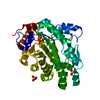
| ||||||||||||
|---|---|---|---|---|---|---|---|---|---|---|---|---|---|
| 1 | 
| ||||||||||||
| Unit cell |
| ||||||||||||
| Components on special symmetry positions |
|
-Components
| #1: Protein | Uroporphyrinogen III decarboxylase / UROD Mass: 39187.254 Da / Num. of mol.: 1 Source method: isolated from a genetically manipulated source Source: (gene. exp.) Nicotiana tabacum (common tobacco) / Production host:  Escherichia coli (E. coli) / References: UniProt: Q42967, uroporphyrinogen decarboxylase Escherichia coli (E. coli) / References: UniProt: Q42967, uroporphyrinogen decarboxylase | ||
|---|---|---|---|
| #2: Chemical | ChemComp-SO4 / Sulfate  Mass: 96.063 Da / Num. of mol.: 5 / Source method: obtained synthetically / Formula: SO4 Mass: 96.063 Da / Num. of mol.: 5 / Source method: obtained synthetically / Formula: SO4#3: Water | ChemComp-HOH / | Water Mass: 18.015 Da / Num. of mol.: 231 / Source method: isolated from a natural source / Formula: H2O Mass: 18.015 Da / Num. of mol.: 231 / Source method: isolated from a natural source / Formula: H2O |
-Experimental details
-Experiment
| Experiment | Method: X-RAY DIFFRACTION / Number of used crystals: 1 |
|---|
- Sample preparation
| Crystal | Density Matthews: 3.14 Å3/Da / Density % sol: 61 % | ||||||||||||||||||||
|---|---|---|---|---|---|---|---|---|---|---|---|---|---|---|---|---|---|---|---|---|---|
| Crystal grow | Temperature: 295 K / Method: vapor diffusion, hanging drop / pH: 9.6 Details: ammonium sulfate, pH 9.6, VAPOR DIFFUSION, HANGING DROP, temperature 295K | ||||||||||||||||||||
| Crystal | *PLUS Density % sol: 61 % | ||||||||||||||||||||
| Crystal grow | *PLUS Temperature: 22 ℃ / pH: 7 | ||||||||||||||||||||
| Components of the solutions | *PLUS
|
-Data collection
| Diffraction | Mean temperature: 100 K |
|---|---|
| Diffraction source | Source: SYNCHROTRON / Site: MPG/DESY, HAMBURG  / Beamline: BW6 / Wavelength: 1.05 Å / Beamline: BW6 / Wavelength: 1.05 Å |
| Detector | Type: MARRESEARCH / Detector: CCD / Date: Mar 2, 1999 |
| Radiation | Protocol: SINGLE WAVELENGTH / Monochromatic (M) / Laue (L): M / Scattering type: x-ray |
| Radiation wavelength | Wavelength: 1.05 Å / Relative weight: 1 |
| Reflection | Resolution: 2.3→18 Å / Num. all: 257709 / Num. obs: 22173 / % possible obs: 97.7 % / Redundancy: 2.14 % / Rsym value: 0.06 / Net I/σ(I): 26.1 |
| Reflection shell | Resolution: 2.3→18 Å / Redundancy: 3.95 % / Mean I/σ(I) obs: 4.3 / Num. unique all: 257709 / Rsym value: 0.346 / % possible all: 98.3 |
| Reflection | *PLUS Num. measured all: 257709 / Rmerge(I) obs: 0.06 |
| Reflection shell | *PLUS Lowest resolution: 2.34 Å / % possible obs: 98.3 % / Rmerge(I) obs: 0.346 |
- Processing
| Software |
| ||||||||||||||||||||
|---|---|---|---|---|---|---|---|---|---|---|---|---|---|---|---|---|---|---|---|---|---|
| Refinement | Method to determine structure: MOLECULAR REPLACEMENT Starting model: PDB entry 1uro Resolution: 2.3→18 Å / Cross valid method: THROUGHOUT / σ(F): 2 / σ(I): 2 / Stereochemistry target values: Engh & Huber / Details: Bulk solvent correction option in CNS
| ||||||||||||||||||||
| Refinement step | Cycle: LAST / Resolution: 2.3→18 Å
| ||||||||||||||||||||
| Refine LS restraints |
| ||||||||||||||||||||
| Xplor file |
| ||||||||||||||||||||
| Software | *PLUS Name: CNS / Classification: refinement | ||||||||||||||||||||
| Refinement | *PLUS σ(F): 2 / % reflection Rfree: 5 % / Rfactor obs: 0.209 | ||||||||||||||||||||
| Solvent computation | *PLUS | ||||||||||||||||||||
| Displacement parameters | *PLUS |
