 Movie
Movie Controller

[English] 日本語
 Yorodumi
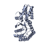
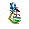
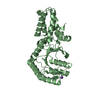
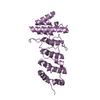
- PDB-1j8m: Signal Recognition Particle conserved GTPase domain from A. ambivalens -
+ Open data

- Basic information
| Entry | Database: PDB / ID: 1j8m | ||||||
|---|---|---|---|---|---|---|---|
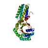
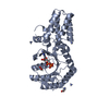
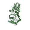
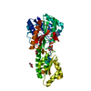
| Title | Signal Recognition Particle conserved GTPase domain from A. ambivalens | ||||||
 Components Components | SIGNAL RECOGNITION 54 KDA PROTEIN | ||||||
 Keywords Keywords |  SIGNALING PROTEIN SIGNALING PROTEIN | ||||||
| Function / homology |  Function and homology informationsignal recognition particle / signal-recognition-particle GTPase / 7S RNA binding / SRP-dependent cotranslational protein targeting to membrane / GTPase activity / GTP binding / ATP hydrolysis activity Function and homology informationsignal recognition particle / signal-recognition-particle GTPase / 7S RNA binding / SRP-dependent cotranslational protein targeting to membrane / GTPase activity / GTP binding / ATP hydrolysis activitySimilarity search - Function | ||||||
| Biological species |  Acidianus ambivalens (archaea) Acidianus ambivalens (archaea) | ||||||
| Method | X-RAY DIFFRACTION / SYNCHROTRON / Resolution: 2 Å | ||||||
 Authors Authors | Montoya, G. / te Kaat, K. / Moll, R. / Schafer, G. / Sinning, I. | ||||||
 Citation Citation | Journal: Structure Fold.Des. / Year: 2000 Title: The crystal structure of the conserved GTPase of SRP54 from the archaeon Acidianus ambivalens and its comparison with related structures suggests a model for the SRP-SRP receptor complex. Authors: Montoya, G. / Kaat, K. / Moll, R. / Schafer, G. / Sinning, I. #1: Journal: Acta Crystallogr.,Sect.D / Year: 1999Title: Crystallization and Preliminary X-ray Diffraction Studies on the Conserved GTPase Domain of the Signal Recognition Particle from Acidianus ambivalens Authors: Montoya, G. / te Kaat, K. / Moll, R. / Schafer, G. / Sinning, I. | ||||||
| History |
|
- Structure visualization
| Structure viewer | Molecule: MolmilJmol/JSmol |
|---|
- Downloads & links
-Download
| PDBx/mmCIF format | 1j8m.cif.gz | 71.9 KB | Display | PDBx/mmCIF format |
|---|---|---|---|---|
| PDB format | pdb1j8m.ent.gz | 54 KB | Display | PDB format |
| PDBx/mmJSON format | 1j8m.json.gz | Tree view | PDBx/mmJSON format | |
| Others |  Other downloads Other downloads |
-Validation report
| Arichive directory | https://data.pdbj.org/pub/pdb/validation_reports/j8/1j8mftp://data.pdbj.org/pub/pdb/validation_reports/j8/1j8m | HTTPS FTP |
|---|
-Related structure data


-Links
 PDBj
PDBj- Assembly
| Deposited unit | 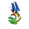
| ||||||||
|---|---|---|---|---|---|---|---|---|---|
| 1 |
| ||||||||
| Unit cell |
|
-Components
| #1: Protein | Mass: 32839.918 Da / Num. of mol.: 1 / Fragment: G-DOMAIN, GTPASE DOMAIN Source method: isolated from a genetically manipulated source Source: (gene. exp.) Acidianus ambivalens (archaea) / Plasmid: pET-16 / Species (production host): Escherichia coli / Production host:  Escherichia coli BL21 (bacteria) / Strain (production host): BL21 / References: UniProt: P70722 Escherichia coli BL21 (bacteria) / Strain (production host): BL21 / References: UniProt: P70722 |
|---|---|
| #2: Water | ChemComp-HOH / Water Mass: 18.015 Da / Num. of mol.: 159 / Source method: isolated from a natural source / Formula: H2O Mass: 18.015 Da / Num. of mol.: 159 / Source method: isolated from a natural source / Formula: H2O |
-Experimental details
-Experiment
| Experiment | Method: X-RAY DIFFRACTION |
|---|
- Sample preparation
| Crystal | Density Matthews: 2.28 Å3/Da / Density % sol: 45.94 % | |||||||||||||||||||||||||
|---|---|---|---|---|---|---|---|---|---|---|---|---|---|---|---|---|---|---|---|---|---|---|---|---|---|---|
| Crystal grow | *PLUS Temperature: 20 ℃ / Method: vapor diffusion, hanging drop | |||||||||||||||||||||||||
| Components of the solutions | *PLUS
|
-Data collection
| Diffraction source | Source: SYNCHROTRON / Site: ESRF  / Beamline: BM14 / Wavelength: 0.932 / Beamline: BM14 / Wavelength: 0.932 |
|---|---|
| Detector | Type: MARRESEARCH / Detector: IMAGE PLATE |
| Radiation | Protocol: SINGLE WAVELENGTH / Monochromatic (M) / Laue (L): M / Scattering type: x-ray |
| Radiation wavelength | Wavelength: 0.932 Å / Relative weight: 1 |
| Reflection | Resolution: 2→40 Å / Num. obs: 20842 |
| Reflection | *PLUS Observed criterion σ(I): 0 / Num. measured all: 451808 / Rmerge(I) obs: 0.051 |
- Processing
| Software |
| ||||||||||||||||||||||||||||||||||||
|---|---|---|---|---|---|---|---|---|---|---|---|---|---|---|---|---|---|---|---|---|---|---|---|---|---|---|---|---|---|---|---|---|---|---|---|---|---|
| Refinement | Resolution: 2→25 Å / SU B: 5.058 / SU ML: 0.145 / SU Rfree: 0.196 / Cross valid method: THROUGHOUT / ESU R: 0.223 / ESU R Free: 0.196
| ||||||||||||||||||||||||||||||||||||
| Displacement parameters | Biso mean: 22.511 Å2
| ||||||||||||||||||||||||||||||||||||
| Refinement step | Cycle: LAST / Resolution: 2→25 Å
| ||||||||||||||||||||||||||||||||||||
| Refine LS restraints |
| ||||||||||||||||||||||||||||||||||||
| LS refinement shell | Resolution: 2→2.052 Å / Total num. of bins used: 20 /
| ||||||||||||||||||||||||||||||||||||
| Software | *PLUS Name: REFMAC / Classification: refinement | ||||||||||||||||||||||||||||||||||||
| Refinement | *PLUS Lowest resolution: 25 Å / % reflection Rfree: 5.1 % | ||||||||||||||||||||||||||||||||||||
| Solvent computation | *PLUS | ||||||||||||||||||||||||||||||||||||
| Displacement parameters | *PLUS | ||||||||||||||||||||||||||||||||||||
| Refine LS restraints | *PLUS
|
