 Movie
Movie Controller


+ Open data

- Basic information
| Entry | Database: PDB / ID: 1j2w | ||||||
|---|---|---|---|---|---|---|---|
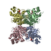
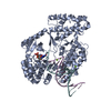
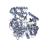
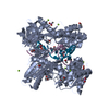
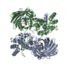
| Title | Tetrameric Structure of aldolase from Thermus thermophilus HB8 | ||||||
 Components Components | Aldolase protein Fructose-bisphosphate aldolase Fructose-bisphosphate aldolase | ||||||
 Keywords Keywords | LYASE / Schiff base / deoxyribose phospahte / carbinolamine / structural genomics / RIKEN Structural Genomics/Proteomics Initiative / RSGI | ||||||
| Function / homology |  Function and homology information Function and homology informationdeoxyribose phosphate catabolic process / deoxyribose-phosphate aldolase / deoxyribose-phosphate aldolase activity / deoxyribonucleotide catabolic process / carbohydrate catabolic process / cytoplasmSimilarity search - Function | ||||||
| Biological species |   Thermus thermophilus (bacteria) Thermus thermophilus (bacteria) | ||||||
| Method | X-RAY DIFFRACTION / SYNCHROTRON / MAD / Resolution: 1.5 Å | ||||||
 Authors Authors | Lokanath, N.K. / Shiromizu, I. / Miyano, M. / Yokoyama, S. / Kuramitsu, S. / Kunishima, N. / RIKEN Structural Genomics/Proteomics Initiative (RSGI) | ||||||
 Citation Citation | Journal: Acta Crystallogr.,Sect.D / Year: 2004 Title: Structure of aldolase from Thermus thermophilus HB8 showing the contribution of oligomeric state to thermostability. Authors: Lokanath, N.K. / Shiromizu, I. / Ohshima, N. / Nodake, Y. / Sugahara, M. / Yokoyama, S. / Kuramitsu, S. / Miyano, M. / Kunishima, N. | ||||||
| History |
|
- Structure visualization
| Structure viewer | Molecule: MolmilJmol/JSmol |
|---|
- Downloads & links
-Download
| PDBx/mmCIF format | 1j2w.cif.gz | 170.3 KB | Display | PDBx/mmCIF format |
|---|---|---|---|---|
| PDB format | pdb1j2w.ent.gz | 137.2 KB | Display | PDB format |
| PDBx/mmJSON format | 1j2w.json.gz | Tree view | PDBx/mmJSON format | |
| Others |  Other downloads Other downloads |
-Validation report
| Arichive directory | https://data.pdbj.org/pub/pdb/validation_reports/j2/1j2wftp://data.pdbj.org/pub/pdb/validation_reports/j2/1j2w | HTTPS FTP |
|---|
-Related structure data


-Links
 PDBj
PDBj
- Assembly
| Deposited unit | 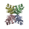
| ||||||||
|---|---|---|---|---|---|---|---|---|---|
| 1 |
| ||||||||
| Unit cell |
|
-Components
| #1: Protein | Fructose-bisphosphate aldolase Mass: 23335.762 Da / Num. of mol.: 4 Source method: isolated from a genetically manipulated source Source: (gene. exp.) Thermus thermophilus (bacteria) / Plasmid: pET-11a / Production host: Escherichia coli (E. coli) / References: UniProt: Q7SIC8, UniProt: Q5SJ28*PLUS#2: Water | ChemComp-HOH / | Water Mass: 18.015 Da / Num. of mol.: 481 / Source method: isolated from a natural source / Formula: H2O Mass: 18.015 Da / Num. of mol.: 481 / Source method: isolated from a natural source / Formula: H2O |
|---|
-Experimental details
-Experiment
| Experiment | Method: X-RAY DIFFRACTION / Number of used crystals: 1 |
|---|
- Sample preparation
| Crystal | Density Matthews: 2.27 Å3/Da / Density % sol: 45.34 % | ||||||||||||||||||||||||||||||
|---|---|---|---|---|---|---|---|---|---|---|---|---|---|---|---|---|---|---|---|---|---|---|---|---|---|---|---|---|---|---|---|
| Crystal grow | Temperature: 295 K / Method: microbatch / pH: 7.9 Details: MPD, Tris HCl, magnesium chloride, pH 7.9, MICROBATCH, temperature 295.0K | ||||||||||||||||||||||||||||||
| Crystal grow | *PLUS pH: 4.9 / Method: batch method | ||||||||||||||||||||||||||||||
| Components of the solutions | *PLUS
|
-Data collection
| Diffraction | Mean temperature: 100 K |
|---|---|
| Diffraction source | Source: SYNCHROTRON / Site: SPring-8  / Beamline: BL26B1 / Wavelength: 0.8 Å / Beamline: BL26B1 / Wavelength: 0.8 Å |
| Detector | Type: RIGAKU RAXIS V / Detector: IMAGE PLATE / Date: Nov 25, 2002 |
| Radiation | Protocol: SINGLE WAVELENGTH / Monochromatic (M) / Laue (L): M / Scattering type: x-ray |
| Radiation wavelength | Wavelength: 0.8 Å / Relative weight: 1 |
| Reflection | Resolution: 1.5→30 Å / Num. all: 137192 / Num. obs: 137075 / % possible obs: 98.3 % / Observed criterion σ(F): 0 / Observed criterion σ(I): 0 / Biso Wilson estimate: 14.62 Å2 |
| Reflection shell | Resolution: 1.5→1.55 Å / % possible all: 99.5 |
| Reflection | *PLUS Highest resolution: 1.5 Å / Num. obs: 137192 / % possible obs: 98.5 % / Redundancy: 4.85 % / Num. measured all: 665297 / Rmerge(I) obs: 0.067 |
| Reflection shell | *PLUS Highest resolution: 1.5 Å / % possible obs: 99.5 % / Rmerge(I) obs: 0.456 / Mean I/σ(I) obs: 4.3 |
- Processing
| Software |
| |||||||||||||||||||||||||
|---|---|---|---|---|---|---|---|---|---|---|---|---|---|---|---|---|---|---|---|---|---|---|---|---|---|---|
| Refinement | Method to determine structure: MAD / Resolution: 1.5→30 Å / Isotropic thermal model: ANISOTROPIC / σ(F): 0 / Stereochemistry target values: Engh & Huber
| |||||||||||||||||||||||||
| Refinement step | Cycle: LAST / Resolution: 1.5→30 Å
| |||||||||||||||||||||||||
| Refine LS restraints |
| |||||||||||||||||||||||||
| Refinement | *PLUS Highest resolution: 1.5 Å / Rfactor Rwork: 0.196 | |||||||||||||||||||||||||
| Solvent computation | *PLUS | |||||||||||||||||||||||||
| Displacement parameters | *PLUS | |||||||||||||||||||||||||
| Refine LS restraints | *PLUS Type: c_angle_deg / Dev ideal: 1.29 |
