 Movie
Movie Controller


+ Open data

- Basic information
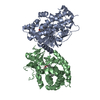
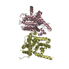
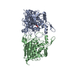
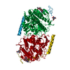
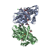
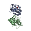
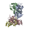
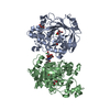
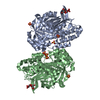
| Entry | Database: PDB / ID: 1ivy | |||||||||
|---|---|---|---|---|---|---|---|---|---|---|
| Title | PHYSIOLOGICAL DIMER HPP PRECURSOR | |||||||||
 Components Components | HUMAN PROTECTIVE PROTEIN Protection Protection | |||||||||
 Keywords Keywords | CARBOXYPEPTIDASE / SERINE CARBOXYPEPTIDASE / PROTECTIVE PROTEIN / GLYCOPROTEIN / ZYMOGEN | |||||||||
| Function / homology |  Function and homology informationcarboxypeptidase C / Defective NEU1 causes sialidosis / serine-type carboxypeptidase activity / Sialic acid metabolism / regulation of chaperone-mediated autophagy / Glycosphingolipid catabolism / negative regulation of chaperone-mediated autophagy / enzyme activator activity / carboxypeptidase activity / MHC class II antigen presentation ...carboxypeptidase C / Defective NEU1 causes sialidosis / serine-type carboxypeptidase activity / Sialic acid metabolism / regulation of chaperone-mediated autophagy / Glycosphingolipid catabolism / negative regulation of chaperone-mediated autophagy / enzyme activator activity / carboxypeptidase activity / MHC class II antigen presentation / lysosomal lumen / intracellular protein transport / regulation of protein stability / azurophil granule lumen / lysosome / intracellular membrane-bounded organelle / Neutrophil degranulation / endoplasmic reticulum / proteolysis / extracellular exosome / extracellular region / membrane Function and homology informationcarboxypeptidase C / Defective NEU1 causes sialidosis / serine-type carboxypeptidase activity / Sialic acid metabolism / regulation of chaperone-mediated autophagy / Glycosphingolipid catabolism / negative regulation of chaperone-mediated autophagy / enzyme activator activity / carboxypeptidase activity / MHC class II antigen presentation ...carboxypeptidase C / Defective NEU1 causes sialidosis / serine-type carboxypeptidase activity / Sialic acid metabolism / regulation of chaperone-mediated autophagy / Glycosphingolipid catabolism / negative regulation of chaperone-mediated autophagy / enzyme activator activity / carboxypeptidase activity / MHC class II antigen presentation / lysosomal lumen / intracellular protein transport / regulation of protein stability / azurophil granule lumen / lysosome / intracellular membrane-bounded organelle / Neutrophil degranulation / endoplasmic reticulum / proteolysis / extracellular exosome / extracellular region / membraneSimilarity search - Function | |||||||||
| Biological species |  Homo sapiens (human) Homo sapiens (human) | |||||||||
| Method | X-RAY DIFFRACTION / SYNCHROTRON / MOLECULAR REPLACEMENT, TWO-FOLD AVERAGING / Resolution: 2.2 Å | |||||||||
 Authors Authors | Rudenko, G. / Bonten, E. / D'Azzo, A. / Hol, W.G.J. | |||||||||
 Citation Citation | Journal: Structure / Year: 1995 Title: Three-dimensional structure of the human 'protective protein': structure of the precursor form suggests a complex activation mechanism. Authors: Rudenko, G. / Bonten, E. / d'Azzo, A. / Hol, W.G. #1: Journal: Acta Crystallogr.,Sect.D / Year: 1996Title: Structure Determination of the Human Protective Protein: Twofold Averaging Reveals the Three-Dimensional Structure of a Domain which Was Entirely Absent in the Initial Model Authors: Rudenko, G. / Bonten, E. / D'Azzo, A. / Hol, W.G.J. #2: Journal: Cell(Cambridge,Mass.) / Year: 1988Title: Expression of Cdna Encoding the Human 'Protective Protein' Associated with Lysosomal Beta-Galactosidase and Neuraminidase: Homology to Yeast Proteases Authors: Galjart, N.J. / Gillemans, N. / Harris, A. / Van Der Horst, G.T. / Verheijen, F.W. / Galjaard, H. / D'Azzo, A. | |||||||||
| History |
|
- Structure visualization
| Structure viewer | Molecule: MolmilJmol/JSmol |
|---|
- Downloads & links
-Download
| PDBx/mmCIF format | 1ivy.cif.gz | 195.9 KB | Display | PDBx/mmCIF format |
|---|---|---|---|---|
| PDB format | pdb1ivy.ent.gz | 155.3 KB | Display | PDB format |
| PDBx/mmJSON format | 1ivy.json.gz | Tree view | PDBx/mmJSON format | |
| Others |  Other downloads Other downloads |
-Validation report
| Arichive directory | https://data.pdbj.org/pub/pdb/validation_reports/iv/1ivyftp://data.pdbj.org/pub/pdb/validation_reports/iv/1ivy | HTTPS FTP |
|---|
-Related structure data
| Similar structure data |
|---|

-Links
 PDBj
PDBj


- Assembly
| Deposited unit | 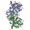
| ||||||||
|---|---|---|---|---|---|---|---|---|---|
| 1 |
| ||||||||
| Unit cell |
|
-Components
| #1: Protein | Protection / PROTECTIVE PROTEIN/CATHEPSIN A / CARBOXYPEPTIDASE L Mass: 51486.004 Da / Num. of mol.: 2 Source method: isolated from a genetically manipulated source Source: (gene. exp.) Homo sapiens (human)Description: BACULOVIRUS MEDIATED OVER-EXPRESSION. SEE BONTEN ET AL. 1995, J.B.C. 270, P. 26441-26445 Cell line: SF21 / Gene: HUPP54 / Gene (production host): HUPP54 / Production host: SF21 INSECT CELLS / References: UniProt: P10619, carboxypeptidase C#2: Polysaccharide | 2-acetamido-2-deoxy-alpha-D-glucopyranose-(1-4)-2-acetamido-2-deoxy-beta-D-glucopyranose | / Mass: 424.401 Da / Num. of mol.: 1Source method: isolated from a genetically manipulated source #3: Polysaccharide | 2-acetamido-2-deoxy-beta-D-glucopyranose-(1-4)-2-acetamido-2-deoxy-beta-D-glucopyranose | / Mass: 424.401 Da / Num. of mol.: 1Source method: isolated from a genetically manipulated source #4: Sugar | N-Acetylglucosamine  Type: D-saccharide, beta linking / Mass: 221.208 Da / Num. of mol.: 2 Type: D-saccharide, beta linking / Mass: 221.208 Da / Num. of mol.: 2Source method: isolated from a genetically manipulated source Formula: C8H15NO6 #5: Water | ChemComp-HOH / | Water Mass: 18.015 Da / Num. of mol.: 296 / Source method: isolated from a natural source / Formula: H2O Mass: 18.015 Da / Num. of mol.: 296 / Source method: isolated from a natural source / Formula: H2O |
|---|
-Experimental details
-Experiment
| Experiment | Method: X-RAY DIFFRACTION / Number of used crystals: 1 |
|---|
- Sample preparation
| Crystal | Density Matthews: 3.2 Å3/Da / Density % sol: 62 % |
|---|---|
| Crystal grow | Temperature: 285 K / pH: 8.3 Details: PEG 8000, PH 8.0 - 8.3 AT 4 - 12 DEGREES CELSIUS, temperature 285K |
| Crystal grow | *PLUS Method: unknown / PH range low: 8.3 / PH range high: 8 |
| Components of the solutions | *PLUS Common name: PEG8000 |
-Data collection
| Diffraction | Mean temperature: 95 K |
|---|---|
| Diffraction source | Source: SYNCHROTRON / Site: SSRL  / Beamline: BL7-1 / Wavelength: 1.08 / Beamline: BL7-1 / Wavelength: 1.08 |
| Detector | Type: MARRESEARCH / Detector: IMAGE PLATE / Date: Mar 24, 1994 |
| Radiation | Monochromatic (M) / Laue (L): M / Scattering type: x-ray |
| Radiation wavelength | Wavelength: 1.08 Å / Relative weight: 1 |
| Reflection | Resolution: 2.2→32.3 Å / Num. obs: 67740 / % possible obs: 95.7 % / Redundancy: 6 % / Rmerge(I) obs: 0.051 / Net I/σ(I): 9.8 |
| Reflection shell | Resolution: 2.2→2.26 Å / Redundancy: 6 % / Mean I/σ(I) obs: 5.7 / Rsym value: 0.13 / % possible all: 87 |
| Reflection | *PLUS Num. measured all: 436709 |
| Reflection shell | *PLUS % possible obs: 87 % / Rmerge(I) obs: 0.13 |
- Processing
| Software |
| ||||||||||||||||||||||||||||||||||||||||||||||||||||||||||||
|---|---|---|---|---|---|---|---|---|---|---|---|---|---|---|---|---|---|---|---|---|---|---|---|---|---|---|---|---|---|---|---|---|---|---|---|---|---|---|---|---|---|---|---|---|---|---|---|---|---|---|---|---|---|---|---|---|---|---|---|---|---|
| Refinement | Method to determine structure: MOLECULAR REPLACEMENT, TWO-FOLD AVERAGING Starting model: SERINE CARBOXYPEPTIDASE FROM WHEAT Resolution: 2.2→8 Å Details: RESIDUES ASN B 216 AND LEU B 217 ARE IN AN AREA OF POOR ELECTRON DENSITY. THE EXACT GEOMETRY OF THESE RESIDUES IS NOT CLEAR FROM THE ELECTRON DENSITY MAP, BUT THEIR GENERAL POSITION IS. IN ...Details: RESIDUES ASN B 216 AND LEU B 217 ARE IN AN AREA OF POOR ELECTRON DENSITY. THE EXACT GEOMETRY OF THESE RESIDUES IS NOT CLEAR FROM THE ELECTRON DENSITY MAP, BUT THEIR GENERAL POSITION IS. IN ADDITION, THE RESTRAINING DISULFIDE CYS 213 - CYS 218 SUPPORTS THE DEPOSITORS' MODEL IN THIS AREA.
| ||||||||||||||||||||||||||||||||||||||||||||||||||||||||||||
| Refine analyze | Luzzati sigma a obs: 0.28 Å | ||||||||||||||||||||||||||||||||||||||||||||||||||||||||||||
| Refinement step | Cycle: LAST / Resolution: 2.2→8 Å
| ||||||||||||||||||||||||||||||||||||||||||||||||||||||||||||
| Refine LS restraints |
| ||||||||||||||||||||||||||||||||||||||||||||||||||||||||||||
| LS refinement shell | Resolution: 2.2→2.3 Å /
| ||||||||||||||||||||||||||||||||||||||||||||||||||||||||||||
| Software | *PLUS Name: X-PLOR / Classification: refinement | ||||||||||||||||||||||||||||||||||||||||||||||||||||||||||||
| Refinement | *PLUS | ||||||||||||||||||||||||||||||||||||||||||||||||||||||||||||
| Solvent computation | *PLUS | ||||||||||||||||||||||||||||||||||||||||||||||||||||||||||||
| Displacement parameters | *PLUS | ||||||||||||||||||||||||||||||||||||||||||||||||||||||||||||
| LS refinement shell | *PLUS Total num. of bins used: 8 / Rfactor obs: 0.24 |
