 Movie
Movie Controller


+ Open data

- Basic information
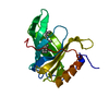
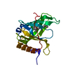
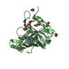
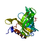
| Entry | Database: PDB / ID: 1iiu | ||||||
|---|---|---|---|---|---|---|---|
| Title | Chicken plasma retinol-binding protein (RBP) | ||||||
 Components Components | plasma retinol-binding protein | ||||||
 Keywords Keywords |  TRANSPORT PROTEIN / RBP / retinol TRANSPORT PROTEIN / RBP / retinol | ||||||
| Function / homology |  Function and homology information Function and homology informationyolk plasma / The canonical retinoid cycle in rods (twilight vision) / Retinoid metabolism and transport / oocyte growth / yolk / retinol transport / retinol transmembrane transporter activity / maintenance of gastrointestinal epithelium / eye development / retinal binding ...yolk plasma / The canonical retinoid cycle in rods (twilight vision) / Retinoid metabolism and transport / oocyte growth / yolk / retinol transport / retinol transmembrane transporter activity / maintenance of gastrointestinal epithelium / eye development / retinal binding / response to vitamin A / retinol metabolic process / clathrin-coated vesicle / retinol binding / response to retinoic acid / gluconeogenesis / response to virus / positive regulation of insulin secretion / response to estrogen / glucose homeostasis / protein-containing complex assembly / protein domain specific binding / signaling receptor binding / extracellular space / extracellular exosomeSimilarity search - Function | ||||||
| Biological species |  Gallus gallus (chicken) Gallus gallus (chicken) | ||||||
| Method | X-RAY DIFFRACTION / MOLECULAR REPLACEMENT / Resolution: 2.5 Å | ||||||
 Authors Authors | Zanotti, G. / Calderone, V. / Berni, R. | ||||||
 Citation Citation | Journal: BIOCHIM.BIOPHYS.ACTA / Year: 2001 Title: Structure of Chicken plasma retinol-binding protein Authors: Zanotti, G. / Calderone, V. / Beda, M. / Malpeli, G. / Folli, C. / Berni, R. #1: Journal: J.Mol.Biol. / Year: 2001Title: Crystal Structure of the Trigonal Form of Human Plasma Retinol-binding Protein at 2.5 A Resolution Authors: Zanotti, G. / Ottonello, S. / Berni, R. / Monaco, H.L. #2: Journal: Eur.J.Biochem. / Year: 1990Title: The bovine plasma retinol-binding protein. Amino acid sequence, interaction with transthyretin, crystallization and preliminary X-ray data. Authors: Berni, R. / Stoppini, M. / Zapponi, M.C. / Meloni, M.L. / Monaco, H.L. / Zanotti, G. #3: Journal: J.Biol.Chem. / Year: 1993Title: Crystal structure of liganded and unliganded forms of bovine plasma retinol-binding protein Authors: Zanotti, G. / Berni, R. / Monaco, H.L. #4: Journal: Proteins / Year: 1990Title: Crystallographic Refinement of Human Serum Retinol Binding Protein at 2 Angstroms Resolution Authors: Cowan, S.W. / Newcomer, M.E. / Jones, T.A. | ||||||
| History |
|
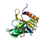
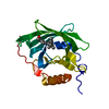
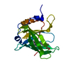
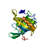
- Structure visualization
| Structure viewer | Molecule: MolmilJmol/JSmol |
|---|
- Downloads & links
-Download
| PDBx/mmCIF format | 1iiu.cif.gz | 48.8 KB | Display | PDBx/mmCIF format |
|---|---|---|---|---|
| PDB format | pdb1iiu.ent.gz | 36.9 KB | Display | PDB format |
| PDBx/mmJSON format | 1iiu.json.gz | Tree view | PDBx/mmJSON format | |
| Others |  Other downloads Other downloads |
-Validation report
| Arichive directory | https://data.pdbj.org/pub/pdb/validation_reports/ii/1iiuftp://data.pdbj.org/pub/pdb/validation_reports/ii/1iiu | HTTPS FTP |
|---|
-Related structure data
| Related structure data | |
|---|---|
| Similar structure data |


-Links
 PDBj
PDBj

- Assembly
| Deposited unit | 
| ||||||||
|---|---|---|---|---|---|---|---|---|---|
| 1 |
| ||||||||
| Unit cell |
|
-Components
| #1: Protein | Mass: 20105.535 Da / Num. of mol.: 1 / Source method: isolated from a natural source / Source: (natural) Gallus gallus (chicken) / References: UniProt: P41263 |
|---|---|
| #2: Chemical | ChemComp-CD /   Mass: 112.411 Da / Num. of mol.: 1 / Source method: obtained synthetically / Formula: Cd Mass: 112.411 Da / Num. of mol.: 1 / Source method: obtained synthetically / Formula: Cd |
| #3: Chemical | ChemComp-RTL / Retinol  Mass: 286.452 Da / Num. of mol.: 1 / Source method: obtained synthetically / Formula: C20H30O Mass: 286.452 Da / Num. of mol.: 1 / Source method: obtained synthetically / Formula: C20H30O |
| #4: Water | ChemComp-HOH / Water Mass: 18.015 Da / Num. of mol.: 95 / Source method: isolated from a natural source / Formula: H2O Mass: 18.015 Da / Num. of mol.: 95 / Source method: isolated from a natural source / Formula: H2O |
-Experimental details
-Experiment
| Experiment | Method: X-RAY DIFFRACTION / Number of used crystals: 1 |
|---|
- Sample preparation
| Crystal | Density Matthews: 2.25 Å3/Da / Density % sol: 45.37 % | ||||||||||||||||||||||||||||||||||||
|---|---|---|---|---|---|---|---|---|---|---|---|---|---|---|---|---|---|---|---|---|---|---|---|---|---|---|---|---|---|---|---|---|---|---|---|---|---|
| Crystal grow | Temperature: 293 K / Method: vapor diffusion, sitting drop / pH: 7 Details: pH 7, VAPOR DIFFUSION, SITTING DROP, temperature 293K | ||||||||||||||||||||||||||||||||||||
| Crystal grow | *PLUS Temperature: 4 ℃ / pH: 6.6 | ||||||||||||||||||||||||||||||||||||
| Components of the solutions | *PLUS
|
-Data collection
| Diffraction | Mean temperature: 293 K |
|---|---|
| Diffraction source | Source: ROTATING ANODE / Type: BRUKER / Wavelength: 1.5418 |
| Detector | Type: SIEMENS HI-STAR / Detector: AREA DETECTOR |
| Radiation | Monochromator: graphite / Protocol: SINGLE WAVELENGTH / Monochromatic (M) / Laue (L): M / Scattering type: x-ray |
| Radiation wavelength | Wavelength: 1.5418 Å / Relative weight: 1 |
| Reflection | Resolution: 2.5→30 Å / Num. obs: 22515 / % possible obs: 82 % / Observed criterion σ(F): 1 / Biso Wilson estimate: 10.9 Å2 / Rsym value: 0.15 |
| Reflection | *PLUS Lowest resolution: 30 Å / Num. obs: 6664 / % possible obs: 87.8 % / Num. measured all: 22515 / Rmerge(I) obs: 0.12 |
| Reflection shell | *PLUS Highest resolution: 2.5 Å / Lowest resolution: 2.66 Å / % possible obs: 68 % / Num. unique obs: 680 / Rmerge(I) obs: 0.32 |
- Processing
| Software |
| ||||||||||||||||||||||||||||||||||||||||||||
|---|---|---|---|---|---|---|---|---|---|---|---|---|---|---|---|---|---|---|---|---|---|---|---|---|---|---|---|---|---|---|---|---|---|---|---|---|---|---|---|---|---|---|---|---|---|
| Refinement | Method to determine structure: MOLECULAR REPLACEMENT / Resolution: 2.5→30.28 Å / Rfactor Rfree error: 0.013 / Data cutoff high absF: 282522.35 / Data cutoff low absF: 0 / Isotropic thermal model: RESTRAINED / Cross valid method: THROUGHOUT / σ(F): 0
| ||||||||||||||||||||||||||||||||||||||||||||
| Solvent computation | Solvent model: flat model / Bsol: 36.8295 Å2 / ksol: 0.289072 e/Å3 | ||||||||||||||||||||||||||||||||||||||||||||
| Displacement parameters | Biso mean: 21.8 Å2
| ||||||||||||||||||||||||||||||||||||||||||||
| Refine analyze |
| ||||||||||||||||||||||||||||||||||||||||||||
| Refinement step | Cycle: LAST / Resolution: 2.5→30.28 Å
| ||||||||||||||||||||||||||||||||||||||||||||
| Refine LS restraints |
| ||||||||||||||||||||||||||||||||||||||||||||
| LS refinement shell | Resolution: 2.5→2.66 Å / Rfactor Rfree error: 0.045 / Total num. of bins used: 6
| ||||||||||||||||||||||||||||||||||||||||||||
| Xplor file |
| ||||||||||||||||||||||||||||||||||||||||||||
| Software | *PLUS Name: CNX / Version: 2000.1 / Classification: refinement | ||||||||||||||||||||||||||||||||||||||||||||
| Refinement | *PLUS σ(F): 0 / % reflection Rfree: 7.5 % / Rfactor obs: 0.21 / Rfactor Rfree: 0.26 | ||||||||||||||||||||||||||||||||||||||||||||
| Solvent computation | *PLUS | ||||||||||||||||||||||||||||||||||||||||||||
| Displacement parameters | *PLUS Biso mean: 21.8 Å2 | ||||||||||||||||||||||||||||||||||||||||||||
| Refine LS restraints | *PLUS
| ||||||||||||||||||||||||||||||||||||||||||||
| LS refinement shell | *PLUS Rfactor Rfree: 0.337 / % reflection Rfree: 7.5 % / Rfactor Rwork: 0.311 |
