 Movie
Movie Controller


+ Open data

- Basic information
| Entry | Database: PDB / ID: 1ihr | ||||||
|---|---|---|---|---|---|---|---|
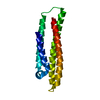
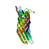
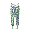
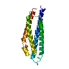
| Title | Crystal structure of the dimeric C-terminal domain of TonB | ||||||
 Components Components | TonB protein | ||||||
 Keywords Keywords |  PROTEIN TRANSPORT / novel fold / intertwined dimer PROTEIN TRANSPORT / novel fold / intertwined dimer | ||||||
| Function / homology |  Function and homology information Function and homology informationreceptor-mediated bacteriophage irreversible attachment to host cell / colicin transport / energy transducer activity / cell envelope / cobalamin transport / siderophore transport / intracellular monoatomic cation homeostasis / plasma membrane protein complex / transmembrane transporter complex / cell outer membrane ...receptor-mediated bacteriophage irreversible attachment to host cell / colicin transport / energy transducer activity / cell envelope / cobalamin transport / siderophore transport / intracellular monoatomic cation homeostasis / plasma membrane protein complex / transmembrane transporter complex / cell outer membrane / transmembrane transport / protein transport / outer membrane-bounded periplasmic space / intracellular iron ion homeostasis / protein domain specific binding / membrane / plasma membraneSimilarity search - Function | ||||||
| Biological species |  Escherichia coli (E. coli) Escherichia coli (E. coli) | ||||||
| Method | X-RAY DIFFRACTION / SYNCHROTRON / MAD / Resolution: 1.55 Å | ||||||
 Authors Authors | Chang, C. / Mooser, A. / Pluckthun, A. / Wlodawer, A. | ||||||
 Citation Citation | Journal: J.Biol.Chem. / Year: 2001 Title: Crystal structure of the dimeric C-terminal domain of TonB reveals a novel fold. Authors: Chang, C. / Mooser, A. / Pluckthun, A. / Wlodawer, A. | ||||||
| History |
|
- Structure visualization
| Structure viewer | Molecule: MolmilJmol/JSmol |
|---|
- Downloads & links
-Download
| PDBx/mmCIF format | 1ihr.cif.gz | 80.8 KB | Display | PDBx/mmCIF format |
|---|---|---|---|---|
| PDB format | pdb1ihr.ent.gz | 61.2 KB | Display | PDB format |
| PDBx/mmJSON format | 1ihr.json.gz | Tree view | PDBx/mmJSON format | |
| Others |  Other downloads Other downloads |
-Validation report
| Arichive directory | https://data.pdbj.org/pub/pdb/validation_reports/ih/1ihrftp://data.pdbj.org/pub/pdb/validation_reports/ih/1ihr | HTTPS FTP |
|---|
-Related structure data
| Similar structure data |
|---|


-Links
 PDBj
PDBj

- Assembly
| Deposited unit | 
| ||||||||
|---|---|---|---|---|---|---|---|---|---|
| 1 |
| ||||||||
| Unit cell |
| ||||||||
| Components on special symmetry positions |
|
-Components
| #1: Protein | Mass: 8563.893 Da / Num. of mol.: 2 / Fragment: C-terminal domain residues 164-239 Source method: isolated from a genetically manipulated source Source: (gene. exp.) Escherichia coli (E. coli) / Gene: JM83 / Plasmid: pAT37 / Production host: Escherichia coli (E. coli) / Strain (production host): XLI-Blue / References: UniProt: P02929#2: Chemical | ChemComp-BR / Bromide  Mass: 79.904 Da / Num. of mol.: 4 / Source method: obtained synthetically / Formula: Br Mass: 79.904 Da / Num. of mol.: 4 / Source method: obtained synthetically / Formula: Br#3: Water | ChemComp-HOH / | Water Mass: 18.015 Da / Num. of mol.: 219 / Source method: isolated from a natural source / Formula: H2O Mass: 18.015 Da / Num. of mol.: 219 / Source method: isolated from a natural source / Formula: H2O |
|---|
-Experimental details
-Experiment
| Experiment | Method: X-RAY DIFFRACTION / Number of used crystals: 1 |
|---|
- Sample preparation
| Crystal | Density Matthews: 1.89 Å3/Da / Density % sol: 35.1 % | ||||||||||||||||||||||||||||||||||||
|---|---|---|---|---|---|---|---|---|---|---|---|---|---|---|---|---|---|---|---|---|---|---|---|---|---|---|---|---|---|---|---|---|---|---|---|---|---|
| Crystal grow | Temperature: 295 K / Method: vapor diffusion, hanging drop / pH: 7.5 Details: PEG 3350, Tris, CaCl2, pH 7.5, VAPOR DIFFUSION, HANGING DROP, temperature 295K | ||||||||||||||||||||||||||||||||||||
| Crystal grow | *PLUS Temperature: 22 ℃ | ||||||||||||||||||||||||||||||||||||
| Components of the solutions | *PLUS
|
-Data collection
| Diffraction | Mean temperature: 100 K | |||||||||||||||
|---|---|---|---|---|---|---|---|---|---|---|---|---|---|---|---|---|
| Diffraction source | Source: SYNCHROTRON / Site: NSLS  / Beamline: X9B / Wavelength: 0.9800, 0.9163, 0.9196, 0.9199 / Beamline: X9B / Wavelength: 0.9800, 0.9163, 0.9196, 0.9199 | |||||||||||||||
| Detector | Type: ADSC QUANTUM 4 / Detector: CCD / Date: Nov 5, 2000 / Details: FOCUSSING MIRROR | |||||||||||||||
| Radiation | Monochromator: SI(111) DOUBLE CRYSTAL / Protocol: MAD / Monochromatic (M) / Laue (L): M / Scattering type: x-ray | |||||||||||||||
| Radiation wavelength |
| |||||||||||||||
| Reflection | Resolution: 1.55→30 Å / Num. all: 22232 / Num. obs: 21518 / % possible obs: 96.8 % / Observed criterion σ(F): -1 / Observed criterion σ(I): -1 / Redundancy: 6.5 % / Biso Wilson estimate: 19.6 Å2 / Rmerge(I) obs: 0.027 / Net I/σ(I): 40.5 | |||||||||||||||
| Reflection shell | Resolution: 1.55→1.6 Å / Redundancy: 2.3 % / Rmerge(I) obs: 0.11 / Mean I/σ(I) obs: 6.8 / Num. unique all: 1745 / % possible all: 80.2 | |||||||||||||||
| Reflection | *PLUS Lowest resolution: 30 Å / Num. measured all: 138987 | |||||||||||||||
| Reflection shell | *PLUS % possible obs: 80.2 % / Rmerge(I) obs: 0.11 |
- Processing
| Software |
| |||||||||||||||||||||||||||||||||
|---|---|---|---|---|---|---|---|---|---|---|---|---|---|---|---|---|---|---|---|---|---|---|---|---|---|---|---|---|---|---|---|---|---|---|
| Refinement | Method to determine structure: MAD / Resolution: 1.55→20 Å / Num. parameters: 12576 / Num. restraintsaints: 14847 / Cross valid method: FREE R / σ(F): 0 / σ(I): 0 / Stereochemistry target values: ENGH AND HUBER
| |||||||||||||||||||||||||||||||||
| Solvent computation | Solvent model: MOEWS & KRETSINGER, J.MOL.BIOL.91(1973)201-228 | |||||||||||||||||||||||||||||||||
| Refine analyze | Luzzati coordinate error obs: 0.16 Å / Luzzati d res low obs: 20 Å / Num. disordered residues: 4 / Occupancy sum hydrogen: 0 / Occupancy sum non hydrogen: 1384.5 | |||||||||||||||||||||||||||||||||
| Refinement step | Cycle: LAST / Resolution: 1.55→20 Å
| |||||||||||||||||||||||||||||||||
| Refine LS restraints |
| |||||||||||||||||||||||||||||||||
| Software | *PLUS Name: SHELXL-97 / Classification: refinement | |||||||||||||||||||||||||||||||||
| Refinement | *PLUS σ(F): 0 / % reflection Rfree: 5.3 % / Rfactor obs: 0.16 / Rfactor Rfree: 0.23 | |||||||||||||||||||||||||||||||||
| Solvent computation | *PLUS | |||||||||||||||||||||||||||||||||
| Displacement parameters | *PLUS |
