 Movie
Movie Controller


+ Open data

- Basic information
| Entry | Database: PDB / ID: 1i4f | ||||||
|---|---|---|---|---|---|---|---|
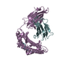
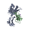
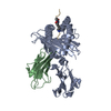
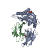
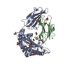
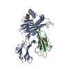
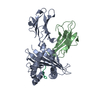
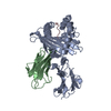
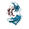
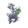
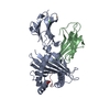
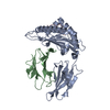
| Title | CRYSTAL STRUCTURE OF HLA-A*0201/MAGE-A4-PEPTIDE COMPLEX | ||||||
 Components Components |
| ||||||
 Keywords Keywords |  IMMUNE SYSTEM / MAJOR HISTOCOMPATIBILITY COMPLEX / HUMAN LEUKOCYTE ANTIGEN / MELANOMA-ASSOCIATED ANTIGEN IMMUNE SYSTEM / MAJOR HISTOCOMPATIBILITY COMPLEX / HUMAN LEUKOCYTE ANTIGEN / MELANOMA-ASSOCIATED ANTIGEN | ||||||
| Function / homology |  Function and homology information Function and homology informationT cell mediated cytotoxicity directed against tumor cell target / antigen processing and presentation of endogenous peptide antigen via MHC class I via ER pathway, TAP-dependent / positive regulation of memory T cell activation / TAP complex binding / antigen processing and presentation of exogenous peptide antigen via MHC class I / Golgi medial cisterna / positive regulation of CD8-positive, alpha-beta T cell activation / CD8-positive, alpha-beta T cell activation / positive regulation of CD8-positive, alpha-beta T cell proliferation / CD8 receptor binding ...T cell mediated cytotoxicity directed against tumor cell target / antigen processing and presentation of endogenous peptide antigen via MHC class I via ER pathway, TAP-dependent / positive regulation of memory T cell activation / TAP complex binding / antigen processing and presentation of exogenous peptide antigen via MHC class I / Golgi medial cisterna / positive regulation of CD8-positive, alpha-beta T cell activation / CD8-positive, alpha-beta T cell activation / positive regulation of CD8-positive, alpha-beta T cell proliferation / CD8 receptor binding / endoplasmic reticulum exit site / beta-2-microglobulin binding / positive regulation of cell cycle / TAP binding / protection from natural killer cell mediated cytotoxicity / antigen processing and presentation of endogenous peptide antigen via MHC class I via ER pathway, TAP-independent / antigen processing and presentation of endogenous peptide antigen via MHC class Ib / detection of bacterium / T cell receptor binding / positive regulation of ferrous iron binding / positive regulation of transferrin receptor binding / early endosome lumen / positive regulation of receptor binding / Nef mediated downregulation of MHC class I complex cell surface expression / DAP12 interactions / negative regulation of receptor binding / lumenal side of endoplasmic reticulum membrane / Endosomal/Vacuolar pathway / Antigen Presentation: Folding, assembly and peptide loading of class I MHC / antigen processing and presentation of exogenous protein antigen via MHC class Ib, TAP-dependent / cellular response to iron(III) ion / negative regulation of forebrain neuron differentiation / ER to Golgi transport vesicle membrane / response to molecule of bacterial origin / regulation of erythrocyte differentiation / regulation of iron ion transport / MHC class I peptide loading complex / HFE-transferrin receptor complex / T cell mediated cytotoxicity / cellular response to iron ion / antigen processing and presentation of endogenous peptide antigen via MHC class I / positive regulation of T cell cytokine production / MHC class I protein complex / multicellular organismal-level iron ion homeostasis / positive regulation of T cell mediated cytotoxicity / peptide antigen assembly with MHC class II protein complex / negative regulation of neurogenesis / positive regulation of receptor-mediated endocytosis / MHC class II protein complex / cellular response to nicotine / histone deacetylase binding / recycling endosome membrane / specific granule lumen / phagocytic vesicle membrane / peptide antigen binding / positive regulation of cellular senescence / antigen processing and presentation of exogenous peptide antigen via MHC class II / Immunoregulatory interactions between a Lymphoid and a non-Lymphoid cell / Interferon gamma signaling / positive regulation of immune response / negative regulation of epithelial cell proliferation / Modulation by Mtb of host immune system / positive regulation of T cell activation / Interferon alpha/beta signaling / positive regulation of type II interferon production / sensory perception of smell / negative regulation of neuron projection development / E3 ubiquitin ligases ubiquitinate target proteins / tertiary granule lumen / DAP12 signaling / MHC class II protein complex binding / late endosome membrane / T cell differentiation in thymus / positive regulation of protein binding / ER-Phagosome pathway / antibacterial humoral response / iron ion transport / T cell receptor signaling pathway / protein refolding / early endosome membrane / protein homotetramerization / intracellular iron ion homeostasis / amyloid fibril formation / learning or memory / defense response to Gram-positive bacterium / immune response / Amyloid fiber formation / lysosomal membrane / endoplasmic reticulum lumen / external side of plasma membrane / Golgi membrane / signaling receptor binding / focal adhesion / innate immune response / Neutrophil degranulation / endoplasmic reticulum membrane / negative regulation of apoptotic process / SARS-CoV-2 activates/modulates innate and adaptive immune responses / Golgi apparatus / negative regulation of transcription by RNA polymerase IISimilarity search - Function | ||||||
| Biological species |  Homo sapiens (human) Homo sapiens (human) | ||||||
| Method | X-RAY DIFFRACTION / SYNCHROTRON / MOLECULAR REPLACEMENT / Resolution: 1.4 Å | ||||||
 Authors Authors | Hillig, R.C. / Coulie, P.G. / Stroobant, V. / Saenger, W. / Ziegler, A. / Huelsmeyer, M. | ||||||
 Citation Citation | Journal: J.Mol.Biol. / Year: 2001 Title: High-resolution structure of HLA-A*0201 in complex with a tumour-specific antigenic peptide encoded by the MAGE-A4 gene. Authors: Hillig, R.C. / Coulie, P.G. / Stroobant, V. / Saenger, W. / Ziegler, A. / Hulsmeyer, M. #1: Journal: Eur.J.Immunol. / Year: 1999Title: A MAGE-A4 Peptide Presented by HLA-A2 is Recognized by Cytolytic T Lymphocytes Authors: Duffour, M.T. / Chaux, P. / Lurquin, C. / Cornelis, G. / Boon, T. / van der Bruggen, P. | ||||||
| History |
|
- Structure visualization
| Structure viewer | Molecule: MolmilJmol/JSmol |
|---|
- Downloads & links
-Download
| PDBx/mmCIF format | 1i4f.cif.gz | 193.2 KB | Display | PDBx/mmCIF format |
|---|---|---|---|---|
| PDB format | pdb1i4f.ent.gz | 152 KB | Display | PDB format |
| PDBx/mmJSON format | 1i4f.json.gz | Tree view | PDBx/mmJSON format | |
| Others |  Other downloads Other downloads |
-Validation report
| Arichive directory | https://data.pdbj.org/pub/pdb/validation_reports/i4/1i4fftp://data.pdbj.org/pub/pdb/validation_reports/i4/1i4f | HTTPS FTP |
|---|
-Related structure data
| Related structure data | 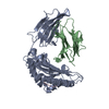 1hhgS S: Starting model for refinement |
|---|---|
| Similar structure data |


-Links
 PDBj
PDBj

- Assembly
| Deposited unit | 
| ||||||||
|---|---|---|---|---|---|---|---|---|---|
| 1 |
| ||||||||
| Unit cell |
| ||||||||
| Details | The biological assembly is a heterotrimeric complex consisting of one HLA-A2 (heavy) chain, one beta-2-microglobulin (light) chain and one MAGE-A4-peptide chain. |
-Components
| #1: Protein | Mass: 31854.203 Da / Num. of mol.: 1 Source method: isolated from a genetically manipulated source Source: (gene. exp.) Homo sapiens (human) / Gene: HLA-A / Production host:  Escherichia coli (E. coli) / Strain (production host): SURE / References: UniProt: P01892, UniProt: P04439*PLUS Escherichia coli (E. coli) / Strain (production host): SURE / References: UniProt: P01892, UniProt: P04439*PLUS | ||
|---|---|---|---|
| #2: Protein | Beta-2 microglobulin Mass: 11879.356 Da / Num. of mol.: 1 Source method: isolated from a genetically manipulated source Source: (gene. exp.) Homo sapiens (human) / Gene: B2M / Production host: Escherichia coli (E. coli) / Strain (production host): SURE / References: UniProt: P61769 | ||
| #3: Protein/peptide | / MAGE-4 ANTIGEN Mass: 1134.200 Da / Num. of mol.: 1 / Fragment: RESIDUES 230-239 / Source method: obtained synthetically Details: This peptide was chemically synthesized. The sequence of the peptide is naturally found in Homo sapiens (humans). References: UniProt: P43358 | ||
| #4: Chemical | Polyethylene glycol  Mass: 252.305 Da / Num. of mol.: 2 / Source method: obtained synthetically / Formula: C11H24O6 Mass: 252.305 Da / Num. of mol.: 2 / Source method: obtained synthetically / Formula: C11H24O6#5: Water | ChemComp-HOH / | Water Mass: 18.015 Da / Num. of mol.: 432 / Source method: isolated from a natural source / Formula: H2O Mass: 18.015 Da / Num. of mol.: 432 / Source method: isolated from a natural source / Formula: H2O |
-Experimental details
-Experiment
| Experiment | Method: X-RAY DIFFRACTION / Number of used crystals: 1 |
|---|
- Sample preparation
| Crystal | Density Matthews: 1.9 Å3/Da / Density % sol: 35.9 % | ||||||||||||||||||||||||||||||||||||
|---|---|---|---|---|---|---|---|---|---|---|---|---|---|---|---|---|---|---|---|---|---|---|---|---|---|---|---|---|---|---|---|---|---|---|---|---|---|
| Crystal grow | Temperature: 291 K / Method: vapor diffusion, hanging drop / pH: 8.5 Details: PEG 8000, Tris-HCl, NaCl. PEG 400 as cryo protectant., pH 8.5, VAPOR DIFFUSION, HANGING DROP, temperature 291K | ||||||||||||||||||||||||||||||||||||
| Crystal grow | *PLUS pH: 7.5 / Details: used microseeding | ||||||||||||||||||||||||||||||||||||
| Components of the solutions | *PLUS
|
-Data collection
| Diffraction | Mean temperature: 100 K |
|---|---|
| Diffraction source | Source: SYNCHROTRON / Site: ESRF  / Beamline: ID13 / Wavelength: 0.782 Å / Beamline: ID13 / Wavelength: 0.782 Å |
| Detector | Type: MARRESEARCH / Detector: CCD / Date: Feb 21, 2000 |
| Radiation | Protocol: SINGLE WAVELENGTH / Monochromatic (M) / Laue (L): M / Scattering type: x-ray |
| Radiation wavelength | Wavelength: 0.782 Å / Relative weight: 1 |
| Reflection | Resolution: 1.4→19.9 Å / Num. all: 324813 / Num. obs: 75194 / % possible obs: 93.7 % / Observed criterion σ(F): 0 / Observed criterion σ(I): 0 / Redundancy: 4.3 % / Biso Wilson estimate: 15.25 Å2 / Rsym value: 0.084 / Net I/σ(I): 15.2 |
| Reflection shell | Resolution: 1.4→1.44 Å / Redundancy: 2.4 % / Mean I/σ(I) obs: 2.3 / Num. unique all: 4563 / Rsym value: 0.369 / % possible all: 80.1 |
| Reflection | *PLUS Rmerge(I) obs: 0.084 |
| Reflection shell | *PLUS % possible obs: 80.1 % / Rmerge(I) obs: 0.369 |
- Processing
| Software |
| |||||||||||||||||||||||||||||||||||||||||||||||||||||||||||||||||||||||||||||||||||||||||||||||||||||||||||||||||||||||||||||||||||||||||||||||||||
|---|---|---|---|---|---|---|---|---|---|---|---|---|---|---|---|---|---|---|---|---|---|---|---|---|---|---|---|---|---|---|---|---|---|---|---|---|---|---|---|---|---|---|---|---|---|---|---|---|---|---|---|---|---|---|---|---|---|---|---|---|---|---|---|---|---|---|---|---|---|---|---|---|---|---|---|---|---|---|---|---|---|---|---|---|---|---|---|---|---|---|---|---|---|---|---|---|---|---|---|---|---|---|---|---|---|---|---|---|---|---|---|---|---|---|---|---|---|---|---|---|---|---|---|---|---|---|---|---|---|---|---|---|---|---|---|---|---|---|---|---|---|---|---|---|---|---|---|---|
| Refinement | Method to determine structure: MOLECULAR REPLACEMENT Starting model: PDB ENTRY 1hhg Resolution: 1.4→19.9 Å / SU B: 2.42983 / SU ML: 0.04954 / Isotropic thermal model: Anisotropic / Cross valid method: THROUGHOUT / σ(F): 0 / ESU R: 0.09996 / ESU R Free: 0.07691 / Stereochemistry target values: Engh & Huber
| |||||||||||||||||||||||||||||||||||||||||||||||||||||||||||||||||||||||||||||||||||||||||||||||||||||||||||||||||||||||||||||||||||||||||||||||||||
| Displacement parameters | Biso mean: 17.26 Å2
| |||||||||||||||||||||||||||||||||||||||||||||||||||||||||||||||||||||||||||||||||||||||||||||||||||||||||||||||||||||||||||||||||||||||||||||||||||
| Refine analyze |
| |||||||||||||||||||||||||||||||||||||||||||||||||||||||||||||||||||||||||||||||||||||||||||||||||||||||||||||||||||||||||||||||||||||||||||||||||||
| Refinement step | Cycle: LAST / Resolution: 1.4→19.9 Å
| |||||||||||||||||||||||||||||||||||||||||||||||||||||||||||||||||||||||||||||||||||||||||||||||||||||||||||||||||||||||||||||||||||||||||||||||||||
| Refine LS restraints |
| |||||||||||||||||||||||||||||||||||||||||||||||||||||||||||||||||||||||||||||||||||||||||||||||||||||||||||||||||||||||||||||||||||||||||||||||||||
| LS refinement shell |
| |||||||||||||||||||||||||||||||||||||||||||||||||||||||||||||||||||||||||||||||||||||||||||||||||||||||||||||||||||||||||||||||||||||||||||||||||||
| Software | *PLUS Name: REFMAC / Classification: refinement | |||||||||||||||||||||||||||||||||||||||||||||||||||||||||||||||||||||||||||||||||||||||||||||||||||||||||||||||||||||||||||||||||||||||||||||||||||
| Refinement | *PLUS Highest resolution: 1.4 Å / σ(F): 0 / % reflection Rfree: 5 % / Rfactor all: 0.138 / Rfactor obs: 0.136 | |||||||||||||||||||||||||||||||||||||||||||||||||||||||||||||||||||||||||||||||||||||||||||||||||||||||||||||||||||||||||||||||||||||||||||||||||||
| Solvent computation | *PLUS | |||||||||||||||||||||||||||||||||||||||||||||||||||||||||||||||||||||||||||||||||||||||||||||||||||||||||||||||||||||||||||||||||||||||||||||||||||
| Displacement parameters | *PLUS | |||||||||||||||||||||||||||||||||||||||||||||||||||||||||||||||||||||||||||||||||||||||||||||||||||||||||||||||||||||||||||||||||||||||||||||||||||
| Refine LS restraints | *PLUS
|
