 Movie
Movie Controller

[English] 日本語
 Yorodumi
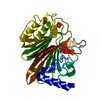
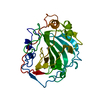
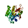
- PDB-1hzg: CRYSTAL STRUCTURE OF THE INACTIVE C866S MUTANT OF THE CATALYTIC D... -
+ Open data

- Basic information
| Entry | Database: PDB / ID: 1hzg | ||||||
|---|---|---|---|---|---|---|---|
| Title | CRYSTAL STRUCTURE OF THE INACTIVE C866S MUTANT OF THE CATALYTIC DOMAIN OF E. COLI CYTOTOXIC NECROTIZING FACTOR 1 | ||||||
 Components Components | CYTOTOXIC NECROTIZING FACTOR 1 | ||||||
 Keywords Keywords |  TOXIN / Beta Sandwich / Rho Deamidase / Rho Transglutaminase TOXIN / Beta Sandwich / Rho Deamidase / Rho Transglutaminase | ||||||
| Function / homology |  Function and homology information Function and homology informationCytotoxic necrotizing factor 1 (CNF1) / Cytotoxic necrotizing factor, Rho-activating domain / Cytotoxic necrotizing factor, Rho-activating domain / Domain of unknown function DUF4765 / Cytotoxic necrotizing factor, Rho-activating domain superfamily / Rho-activating domain of cytotoxic necrotizing factor / Domain of unknown function (DUF4765) / Domain of unknown function DUF6543 / Family of unknown function (DUF6543) / Cytotoxic necrotizing factor-like, catalytic ...Cytotoxic necrotizing factor 1 (CNF1) / Cytotoxic necrotizing factor, Rho-activating domain / Cytotoxic necrotizing factor, Rho-activating domain / Domain of unknown function DUF4765 / Cytotoxic necrotizing factor, Rho-activating domain superfamily / Rho-activating domain of cytotoxic necrotizing factor / Domain of unknown function (DUF4765) / Domain of unknown function DUF6543 / Family of unknown function (DUF6543) / Cytotoxic necrotizing factor-like, catalytic / 4-Layer Sandwich / Alpha Beta Similarity search - Domain/homology | ||||||
| Biological species |  Escherichia coli (E. coli) Escherichia coli (E. coli) | ||||||
| Method | X-RAY DIFFRACTION / Difference Fourier / Resolution: 1.86 Å | ||||||
 Authors Authors | Buetow, L. / Flatau, G. / Chiu, K. / Boquet, P. / Ghosh, P. | ||||||
 Citation Citation | Journal: Nat.Struct.Biol. / Year: 2001 Title: Structure of the Rho-activating domain of Escherichia coli cytotoxic necrotizing factor 1. Authors: Buetow, L. / Flatau, G. / Chiu, K. / Boquet, P. / Ghosh, P. | ||||||
| History |
|
- Structure visualization
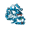
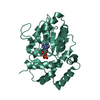
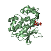
| Structure viewer | Molecule: MolmilJmol/JSmol |
|---|
- Downloads & links
-Download
| PDBx/mmCIF format | 1hzg.cif.gz | 74.2 KB | Display | PDBx/mmCIF format |
|---|---|---|---|---|
| PDB format | pdb1hzg.ent.gz | 53.8 KB | Display | PDB format |
| PDBx/mmJSON format | 1hzg.json.gz | Tree view | PDBx/mmJSON format | |
| Others |  Other downloads Other downloads |
-Validation report
| Arichive directory | https://data.pdbj.org/pub/pdb/validation_reports/hz/1hzgftp://data.pdbj.org/pub/pdb/validation_reports/hz/1hzg | HTTPS FTP |
|---|
-Related structure data
| Related structure data |  1hq0SC S: Starting model for refinement C: citing same article ( |
|---|---|
| Similar structure data |


-Links
 PDBj
PDBj- Assembly
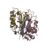
| Deposited unit | 
| ||||||||
|---|---|---|---|---|---|---|---|---|---|
| 1 |
| ||||||||
| Unit cell |
|
-Components
| #1: Protein | Mass: 32642.375 Da / Num. of mol.: 1 / Fragment: CATALYTIC DOMAIN / Mutation: C866S, L794P Source method: isolated from a genetically manipulated source Source: (gene. exp.) Escherichia coli (E. coli) / Strain: E-B35 / Plasmid: PET 28A / Species (production host): Escherichia coli / Production host: Escherichia coli BL21(DE3) (bacteria) / Strain (production host): BL21(DE3) / References: UniProt: Q47106 | ||
|---|---|---|---|
| #2: Chemical | Phosphate  Mass: 94.971 Da / Num. of mol.: 2 / Source method: obtained synthetically / Formula: PO4 Mass: 94.971 Da / Num. of mol.: 2 / Source method: obtained synthetically / Formula: PO4#3: Water | ChemComp-HOH / | Water Mass: 18.015 Da / Num. of mol.: 157 / Source method: isolated from a natural source / Formula: H2O Mass: 18.015 Da / Num. of mol.: 157 / Source method: isolated from a natural source / Formula: H2O |
-Experimental details
-Experiment
| Experiment | Method: X-RAY DIFFRACTION / Number of used crystals: 1 |
|---|
- Sample preparation
| Crystal | Density Matthews: 2.36 Å3/Da / Density % sol: 47.78 % | ||||||||||||||||||||||||||||||||||||||||||||||||||||||||||||
|---|---|---|---|---|---|---|---|---|---|---|---|---|---|---|---|---|---|---|---|---|---|---|---|---|---|---|---|---|---|---|---|---|---|---|---|---|---|---|---|---|---|---|---|---|---|---|---|---|---|---|---|---|---|---|---|---|---|---|---|---|---|
| Crystal grow | Temperature: 310 K / Method: vapor diffusion, hanging drop / pH: 7.5 Details: sodium phosphate, potassium phosphate, 1,4-butandiol, HEPES, DTT, pH 7.5, VAPOR DIFFUSION, HANGING DROP, temperature 310K | ||||||||||||||||||||||||||||||||||||||||||||||||||||||||||||
| Crystal grow | *PLUS Temperature: 37 ℃ / pH: 8 / Method: vapor diffusion | ||||||||||||||||||||||||||||||||||||||||||||||||||||||||||||
| Components of the solutions | *PLUS
|
-Data collection
| Diffraction | Mean temperature: 110 K |
|---|---|
| Diffraction source | Source: ROTATING ANODE / Type: RIGAKU FR-5 / Wavelength: 1.5418 Å |
| Detector | Type: MARRESEARCH / Detector: IMAGE PLATE / Date: Aug 4, 2000 / Details: Supper mirror |
| Radiation | Monochromator: Supper mirror / Protocol: SINGLE WAVELENGTH / Monochromatic (M) / Laue (L): M / Scattering type: x-ray |
| Radiation wavelength | Wavelength: 1.5418 Å / Relative weight: 1 |
| Reflection | Resolution: 1.86→15 Å / Num. all: 25165 / Num. obs: 25142 / % possible obs: 99.9 % / Observed criterion σ(F): 0 / Observed criterion σ(I): -3 / Redundancy: 10 % / Biso Wilson estimate: 20.4 Å2 / Rmerge(I) obs: 0.079 / Net I/σ(I): 33.8 |
| Reflection shell | Resolution: 1.86→1.91 Å / Redundancy: 8.6 % / Rmerge(I) obs: 0.752 / Mean I/σ(I) obs: 3.5 / % possible all: 98.1 |
| Reflection | *PLUS Lowest resolution: 15 Å |
- Processing
| Software |
| ||||||||||||||||||||||||||||||||||||||||
|---|---|---|---|---|---|---|---|---|---|---|---|---|---|---|---|---|---|---|---|---|---|---|---|---|---|---|---|---|---|---|---|---|---|---|---|---|---|---|---|---|---|
| Refinement | Method to determine structure: Difference Fourier Starting model: PDB Entry 1HQ0 Resolution: 1.86→15 Å / Rfactor Rfree error: 0.006 / Data cutoff high absF: 1444667.46 / Data cutoff low absF: 0 / Isotropic thermal model: RESTRAINED / Cross valid method: THROUGHOUT / σ(F): 0 / σ(I): 0 / Stereochemistry target values: Engh & Huber / Details: Used Maximum Likelihood Refinement
| ||||||||||||||||||||||||||||||||||||||||
| Solvent computation | Solvent model: FLAT MODEL / Bsol: 49.38 Å2 / ksol: 0.351 e/Å3 | ||||||||||||||||||||||||||||||||||||||||
| Displacement parameters | Biso mean: 30.1 Å2
| ||||||||||||||||||||||||||||||||||||||||
| Refine analyze |
| ||||||||||||||||||||||||||||||||||||||||
| Refinement step | Cycle: LAST / Resolution: 1.86→15 Å
| ||||||||||||||||||||||||||||||||||||||||
| Refine LS restraints |
| ||||||||||||||||||||||||||||||||||||||||
| LS refinement shell | Resolution: 1.86→1.98 Å / Rfactor Rfree error: 0.022 / Total num. of bins used: 6
| ||||||||||||||||||||||||||||||||||||||||
| Xplor file |
| ||||||||||||||||||||||||||||||||||||||||
| Software | *PLUS Name: CNS / Version: 1 / Classification: refinement | ||||||||||||||||||||||||||||||||||||||||
| Refinement | *PLUS Lowest resolution: 15 Å / σ(F): 0 / % reflection Rfree: 4.9 % / Rfactor obs: 0.198 / Rfactor Rfree: 0.22 | ||||||||||||||||||||||||||||||||||||||||
| Solvent computation | *PLUS | ||||||||||||||||||||||||||||||||||||||||
| Displacement parameters | *PLUS | ||||||||||||||||||||||||||||||||||||||||
| Refine LS restraints | *PLUS
| ||||||||||||||||||||||||||||||||||||||||
| LS refinement shell | *PLUS Rfactor Rfree: 0.288 / % reflection Rfree: 4 % / Rfactor Rwork: 0.258 |
