 Movie
Movie Controller

[English] 日本語
 Yorodumi
- PDB-1hxi: AN UNEXPECTED EXTENDED CONFORMATION FOR THE THIRD TPR MOTIF OF TH... -
+ Open data

- Basic information
| Entry | Database: PDB / ID: 1hxi | ||||||
|---|---|---|---|---|---|---|---|
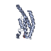
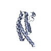
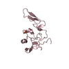
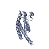
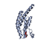
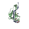
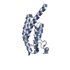
| Title | AN UNEXPECTED EXTENDED CONFORMATION FOR THE THIRD TPR MOTIF OF THE PEROXIN PEX5 FROM TRYPANOSOMA BRUCEI | ||||||
 Components Components | PEROXISOME TARGETING SIGNAL 1 RECEPTOR PEX5 | ||||||
 Keywords Keywords |  TRANSPORT PROTEIN / alpha helical TRANSPORT PROTEIN / alpha helical | ||||||
| Function / homology |  Function and homology information Function and homology informationnucleoside transmembrane transporter activity / peroxisome / membrane => GO:0016020Similarity search - Function | ||||||
| Biological species |  Trypanosoma brucei (eukaryote) Trypanosoma brucei (eukaryote) | ||||||
| Method | X-RAY DIFFRACTION / SYNCHROTRON / MAD / Resolution: 1.6 Å | ||||||
 Authors Authors | Kumar, A. / Roach, C. / Hirsh, I.S. / Turley, S. / deWalque, S. / Michels, P.A.M. / Hol, W.G.J. | ||||||
 Citation Citation | Journal: J.Mol.Biol. / Year: 2001 Title: An unexpected extended conformation for the third TPR motif of the peroxin PEX5 from Trypanosoma brucei. Authors: Kumar, A. / Roach, C. / Hirsh, I.S. / Turley, S. / deWalque, S. / Michels, P.A. / Hol, W.G. | ||||||
| History |
|
- Structure visualization
| Structure viewer | Molecule: MolmilJmol/JSmol |
|---|
- Downloads & links
-Download
| PDBx/mmCIF format | 1hxi.cif.gz | 33.7 KB | Display | PDBx/mmCIF format |
|---|---|---|---|---|
| PDB format | pdb1hxi.ent.gz | 25.9 KB | Display | PDB format |
| PDBx/mmJSON format | 1hxi.json.gz | Tree view | PDBx/mmJSON format | |
| Others |  Other downloads Other downloads |
-Validation report
| Arichive directory | https://data.pdbj.org/pub/pdb/validation_reports/hx/1hxiftp://data.pdbj.org/pub/pdb/validation_reports/hx/1hxi | HTTPS FTP |
|---|
-Related structure data
| Similar structure data |
|---|


-Links
 PDBj
PDBj


- Assembly
| Deposited unit | 
| ||||||||
|---|---|---|---|---|---|---|---|---|---|
| 1 |
| ||||||||
| Unit cell |
|
-Components
| #1: Protein | Mass: 13713.620 Da / Num. of mol.: 1 / Fragment: FIRST THREE TPR MOTIFS Source method: isolated from a genetically manipulated source Source: (gene. exp.) Trypanosoma brucei (eukaryote) / Gene: PEX5 / Plasmid: PET26B / Species (production host): Escherichia coli / Production host:  Escherichia coli BL21(DE3) (bacteria) / Strain (production host): BL21(DE3) / References: UniProt: Q9U763, UniProt: Q9U7C3*PLUS Escherichia coli BL21(DE3) (bacteria) / Strain (production host): BL21(DE3) / References: UniProt: Q9U763, UniProt: Q9U7C3*PLUS |
|---|---|
| #2: Chemical | ChemComp-MG /   Mass: 24.305 Da / Num. of mol.: 1 / Source method: obtained synthetically / Formula: Mg Mass: 24.305 Da / Num. of mol.: 1 / Source method: obtained synthetically / Formula: Mg |
| #3: Water | ChemComp-HOH / Water Mass: 18.015 Da / Num. of mol.: 146 / Source method: isolated from a natural source / Formula: H2O Mass: 18.015 Da / Num. of mol.: 146 / Source method: isolated from a natural source / Formula: H2O |
-Experimental details
-Experiment
| Experiment | Method: X-RAY DIFFRACTION / Number of used crystals: 1 |
|---|
- Sample preparation
| Crystal | Density Matthews: 3.118 Å3/Da / Density % sol: 61.7 % | ||||||||||||||||||||||||||||||
|---|---|---|---|---|---|---|---|---|---|---|---|---|---|---|---|---|---|---|---|---|---|---|---|---|---|---|---|---|---|---|---|
| Crystal grow | Temperature: 298 K / Method: vapor diffusion, sitting drop / pH: 6.5 Details: Imidazole, MgCl2, IPTG, DMSO, pH 6.5, VAPOR DIFFUSION, SITTING DROP, temperature 298K | ||||||||||||||||||||||||||||||
| Crystal grow | *PLUS Temperature: 20 ℃ | ||||||||||||||||||||||||||||||
| Components of the solutions | *PLUS
|
-Data collection
| Diffraction | Mean temperature: 100 K | ||||||||||||
|---|---|---|---|---|---|---|---|---|---|---|---|---|---|
| Diffraction source | Source: SYNCHROTRON / Site: ALS  / Beamline: 5.0.2 / Wavelength: 0.9798, 0.9800, 0.9574 / Beamline: 5.0.2 / Wavelength: 0.9798, 0.9800, 0.9574 | ||||||||||||
| Detector | Type: ADSC QUANTUM 4 / Detector: CCD / Date: Apr 17, 2000 | ||||||||||||
| Radiation | Monochromator: synchrotron / Protocol: MAD / Monochromatic (M) / Laue (L): M / Scattering type: x-ray | ||||||||||||
| Radiation wavelength |
| ||||||||||||
| Reflection | Resolution: 1.6→20 Å / Num. all: 23687 / Num. obs: 23237 / % possible obs: 98.1 % / Observed criterion σ(F): 0 / Observed criterion σ(I): 0 / Redundancy: 5.8 % / Biso Wilson estimate: 20.049 Å2 / Rmerge(I) obs: 0.079 / Net I/σ(I): 17.3 | ||||||||||||
| Reflection shell | Resolution: 1.6→20 Å / Redundancy: 5.6 % / Rmerge(I) obs: 0.502 / % possible all: 99.7 | ||||||||||||
| Reflection shell | *PLUS % possible obs: 99.7 % / Mean I/σ(I) obs: 3.6 |
- Processing
| Software |
| |||||||||||||||||||||||||
|---|---|---|---|---|---|---|---|---|---|---|---|---|---|---|---|---|---|---|---|---|---|---|---|---|---|---|
| Refinement | Method to determine structure: MAD / Resolution: 1.6→20 Å / σ(F): 0 / σ(I): 0 / Stereochemistry target values: REFMAC
| |||||||||||||||||||||||||
| Displacement parameters | Biso mean: 20.5 Å2 | |||||||||||||||||||||||||
| Refinement step | Cycle: LAST / Resolution: 1.6→20 Å
| |||||||||||||||||||||||||
| Refine LS restraints |
| |||||||||||||||||||||||||
| Software | *PLUS Name: REFMAC / Classification: refinement | |||||||||||||||||||||||||
| Refine LS restraints | *PLUS
|
