 Movie
Movie Controller


+ Open data

- Basic information
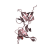
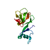
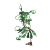
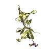
| Entry | Database: PDB / ID: 1hus | ||||||
|---|---|---|---|---|---|---|---|
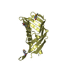
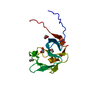
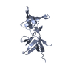
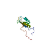
| Title | RIBOSOMAL PROTEIN S7 | ||||||
 Components Components | RIBOSOMAL PROTEIN S7 | ||||||
 Keywords Keywords | RIBOSOMAL PROTEIN / RNA-BINDING PROTEIN / DECODING CENTER | ||||||
| Function / homology |  Function and homology information Function and homology informationsmall ribosomal subunit / tRNA binding / rRNA binding / structural constituent of ribosome / translationSimilarity search - Function | ||||||
| Biological species |   Geobacillus stearothermophilus (bacteria) Geobacillus stearothermophilus (bacteria) | ||||||
| Method | X-RAY DIFFRACTION / SYNCHROTRON / MAD WITH SE-MET MUTANT / Resolution: 2.5 Å | ||||||
 Authors Authors | Hosaka, H. / Nakagawa, A. / Tanaka, I. | ||||||
 Citation Citation | Journal: Structure / Year: 1997 Title: Ribosomal protein S7: a new RNA-binding motif with structural similarities to a DNA architectural factor. Authors: Hosaka, H. / Nakagawa, A. / Tanaka, I. / Harada, N. / Sano, K. / Kimura, M. / Yao, M. / Wakatsuki, S. #1: Journal: J.Struct.Biol. / Year: 1997Title: Crystallization and Preliminary X-Ray Crystallographic Study of the Ribosomal Protein S7 from Bacillus Stearothermophilus Authors: Harada, N. / Sano, K. / Kimura, M. / Hosaka, H. / Nakagawa, A. / Tanaka, I. | ||||||
| History |
|
- Structure visualization
| Structure viewer | Molecule: MolmilJmol/JSmol |
|---|
- Downloads & links
-Download
| PDBx/mmCIF format | 1hus.cif.gz | 36.4 KB | Display | PDBx/mmCIF format |
|---|---|---|---|---|
| PDB format | pdb1hus.ent.gz | 28.6 KB | Display | PDB format |
| PDBx/mmJSON format | 1hus.json.gz | Tree view | PDBx/mmJSON format | |
| Others |  Other downloads Other downloads |
-Validation report
| Arichive directory | https://data.pdbj.org/pub/pdb/validation_reports/hu/1husftp://data.pdbj.org/pub/pdb/validation_reports/hu/1hus | HTTPS FTP |
|---|
-Related structure data
| Similar structure data |
|---|


-Links
 PDBj
PDBj- Assembly
| Deposited unit | 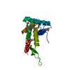
| ||||||||
|---|---|---|---|---|---|---|---|---|---|
| 1 |
| ||||||||
| Unit cell |
|
-Components
| #1: Protein | Mass: 18230.283 Da / Num. of mol.: 1 Source method: isolated from a genetically manipulated source Source: (gene. exp.) Geobacillus stearothermophilus (bacteria)Cell line: BL21 / Gene: S7 / Plasmid: BL21 / Gene (production host): S7 / Production host: Escherichia coli (E. coli) / Strain (production host): BL21 (DE3) LYSS / References: UniProt: P22744 |
|---|---|
| #2: Water | ChemComp-HOH / Water Mass: 18.015 Da / Num. of mol.: 25 / Source method: isolated from a natural source / Formula: H2O Mass: 18.015 Da / Num. of mol.: 25 / Source method: isolated from a natural source / Formula: H2O |
-Experimental details
-Experiment
| Experiment | Method: X-RAY DIFFRACTION / Number of used crystals: 1 |
|---|
- Sample preparation
| Crystal | Density Matthews: 2.9 Å3/Da / Density % sol: 57 % | ||||||||||||||||||||
|---|---|---|---|---|---|---|---|---|---|---|---|---|---|---|---|---|---|---|---|---|---|
| Crystal grow | pH: 8.2 Details: PROTEIN WAS CRYSTALLIZED FROM 0.1M NA HEPES BUFFER(PH8.2) WITH 4%(V/V) 2-PROPANOL AND 2.0M AMMONIUM SULFATE. | ||||||||||||||||||||
| Crystal grow | *PLUS Method: unknown | ||||||||||||||||||||
| Components of the solutions | *PLUS
|
-Data collection
| Diffraction | Mean temperature: 293 K |
|---|---|
| Diffraction source | Source: SYNCHROTRON / Site: ESRF  / Beamline: BM14 / Wavelength: 0.90007 / Beamline: BM14 / Wavelength: 0.90007 |
| Detector | Type: PRINCETON 2K / Detector: CCD / Date: Mar 1, 1997 / Details: COLLIMATING AND FOCUSING MIRRORS |
| Radiation | Monochromator: SI(111) / Monochromatic (M) / Laue (L): M / Scattering type: x-ray |
| Radiation wavelength | Wavelength: 0.90007 Å / Relative weight: 1 |
| Reflection | Resolution: 2.5→30 Å / Num. obs: 7586 / % possible obs: 97.6 % / Observed criterion σ(I): 18.7 / Redundancy: 2.8 % / Biso Wilson estimate: 33.3 Å2 / Rmerge(I) obs: 0.048 / Net I/σ(I): 20.3 |
| Reflection shell | Resolution: 2.5→2.54 Å / Redundancy: 2.2 % / Rmerge(I) obs: 0.138 / Mean I/σ(I) obs: 5.8 / % possible all: 92.9 |
| Reflection | *PLUS Num. measured all: 53236 |
| Reflection shell | *PLUS % possible obs: 92.9 % |
- Processing
| Software |
| ||||||||||||||||||||||||||||||||||||||||||||||||||||||||||||||||||||||||||||||||
|---|---|---|---|---|---|---|---|---|---|---|---|---|---|---|---|---|---|---|---|---|---|---|---|---|---|---|---|---|---|---|---|---|---|---|---|---|---|---|---|---|---|---|---|---|---|---|---|---|---|---|---|---|---|---|---|---|---|---|---|---|---|---|---|---|---|---|---|---|---|---|---|---|---|---|---|---|---|---|---|---|---|
| Refinement | Method to determine structure: MAD WITH SE-MET MUTANT / Resolution: 2.5→8 Å / Rfactor Rfree error: 0.01 / Isotropic thermal model: RESTRAINED / Cross valid method: THROUGHOUT / σ(F): 0 Details: REFMAC (G.N.MURSHUDOV ET AL.) WAS ALSO USED DURING THE REFINEMENT CYCLE. PARAMETERS OF SE-MET WAS DERIVED FROM THOSE OF METHIONINE.
| ||||||||||||||||||||||||||||||||||||||||||||||||||||||||||||||||||||||||||||||||
| Displacement parameters | Biso mean: 33.4 Å2 | ||||||||||||||||||||||||||||||||||||||||||||||||||||||||||||||||||||||||||||||||
| Refine analyze |
| ||||||||||||||||||||||||||||||||||||||||||||||||||||||||||||||||||||||||||||||||
| Refinement step | Cycle: LAST / Resolution: 2.5→8 Å
| ||||||||||||||||||||||||||||||||||||||||||||||||||||||||||||||||||||||||||||||||
| Refine LS restraints |
| ||||||||||||||||||||||||||||||||||||||||||||||||||||||||||||||||||||||||||||||||
| LS refinement shell | Resolution: 2.5→2.61 Å / Rfactor Rfree error: 0.038 / Total num. of bins used: 8
| ||||||||||||||||||||||||||||||||||||||||||||||||||||||||||||||||||||||||||||||||
| Software | *PLUS Name: X-PLOR / Version: 3.851 / Classification: refinement | ||||||||||||||||||||||||||||||||||||||||||||||||||||||||||||||||||||||||||||||||
| Refinement | *PLUS Rfactor Rfree: 0.27 | ||||||||||||||||||||||||||||||||||||||||||||||||||||||||||||||||||||||||||||||||
| Solvent computation | *PLUS | ||||||||||||||||||||||||||||||||||||||||||||||||||||||||||||||||||||||||||||||||
| Displacement parameters | *PLUS | ||||||||||||||||||||||||||||||||||||||||||||||||||||||||||||||||||||||||||||||||
| Refine LS restraints | *PLUS
|
