 Movie
Movie Controller

[English] 日本語
 Yorodumi
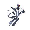
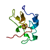
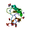
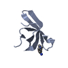
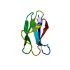
- PDB-1hip: TWO-ANGSTROM CRYSTAL STRUCTURE OF OXIDIZED CHROMATIUM HIGH POTENT... -
+ Open data

- Basic information
| Entry | Database: PDB / ID: 1hip | ||||||
|---|---|---|---|---|---|---|---|
| Title | TWO-ANGSTROM CRYSTAL STRUCTURE OF OXIDIZED CHROMATIUM HIGH POTENTIAL IRON PROTEIN | ||||||
 Components Components | HIGH POTENTIAL IRON PROTEIN | ||||||
 Keywords Keywords | ELECTRON TRANSFER (IRON-SULFUR PROTEIN) | ||||||
| Function / homology |  Function and homology information Function and homology informationaerobic electron transport chain / 4 iron, 4 sulfur cluster binding /  electron transfer activity / periplasmic space / metal ion binding electron transfer activity / periplasmic space / metal ion bindingSimilarity search - Function | ||||||
| Biological species |  Allochromatium vinosum (bacteria) Allochromatium vinosum (bacteria) | ||||||
| Method | X-RAY DIFFRACTION / Resolution: 2 Å | ||||||
 Authors Authors | Carterjunior, C.W. / Kraut, J. / Freer, S.T. / Xuong, N.-H. / Alden, R.A. / Bartsch, R.G. | ||||||
 Citation Citation | Journal: J.Biol.Chem. / Year: 1974 Title: Two-Angstrom crystal structure of oxidized Chromatium high potential iron protein. Authors: Carter Jr., C.W. / Kraut, J. / Freer, S.T. / Xuong, N.H. / Alden, R.A. / Bartsch, R.G. #1: Journal: J.Biol.Chem. / Year: 1975Title: Crystallographic Structure Refinement of Chromatium High Potential Iron Protein at Two Angstroms Resolution Authors: Freer, S.T. / Alden, R.A. / Carterjunior, C.W. / Kraut, J. #2: Journal: J.Biol.Chem. / Year: 1974Title: Comparison of Oxidation-Reduction Site Geometries in Oxidized and Reduced Chromatium High Potential Iron Protein and Oxidized Peptococcus Aerogenes Ferredoxin Authors: Carterjunior, C.W. / Kraut, J. / Freer, S.T. / Alden, R.A. #3: Journal: Cold Spring Harbor Symp.Quant.Biol. / Year: 1972Title: Structure of the Iron-Sulfur Cluster in the Chromatium Iron Protein at 2.25 Angstroms Resolution Authors: Carterjunior, C.W. / Freer, S.T. / Xuong, N.H. / Alden, R.A. / Kraut, J. | ||||||
| History |
|
- Structure visualization
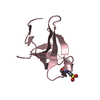
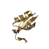
| Structure viewer | Molecule: MolmilJmol/JSmol |
|---|
- Downloads & links
-Download
| PDBx/mmCIF format | 1hip.cif.gz | 26.3 KB | Display | PDBx/mmCIF format |
|---|---|---|---|---|
| PDB format | pdb1hip.ent.gz | 15.7 KB | Display | PDB format |
| PDBx/mmJSON format | 1hip.json.gz | Tree view | PDBx/mmJSON format | |
| Others |  Other downloads Other downloads |
-Validation report
| Arichive directory | https://data.pdbj.org/pub/pdb/validation_reports/hi/1hipftp://data.pdbj.org/pub/pdb/validation_reports/hi/1hip | HTTPS FTP |
|---|
-Related structure data
| Similar structure data |
|---|


-Links
 PDBj
PDBj

- Assembly
| Deposited unit | 
| ||||||||
|---|---|---|---|---|---|---|---|---|---|
| 1 |
| ||||||||
| Unit cell |
|
-Components
| #1: Protein | Mass: 8912.928 Da / Num. of mol.: 1 Source method: isolated from a genetically manipulated source Source: (gene. exp.) Allochromatium vinosum (bacteria) / References: UniProt: P00260 |
|---|---|
| #2: Chemical | ChemComp-SF4 / Iron–sulfur cluster  Mass: 351.640 Da / Num. of mol.: 1 / Source method: obtained synthetically / Formula: Fe4S4 Mass: 351.640 Da / Num. of mol.: 1 / Source method: obtained synthetically / Formula: Fe4S4 |
| #3: Water | ChemComp-HOH / Water Mass: 18.015 Da / Num. of mol.: 75 / Source method: isolated from a natural source / Formula: H2O Mass: 18.015 Da / Num. of mol.: 75 / Source method: isolated from a natural source / Formula: H2O |
-Experimental details
-Experiment
| Experiment | Method: X-RAY DIFFRACTION |
|---|
- Sample preparation
| Crystal | Density Matthews: 1.91 Å3/Da / Density % sol: 35.54 % | ||||||||||||||||||||||||||||
|---|---|---|---|---|---|---|---|---|---|---|---|---|---|---|---|---|---|---|---|---|---|---|---|---|---|---|---|---|---|
| Crystal | *PLUS Density % sol: 30 % | ||||||||||||||||||||||||||||
| Crystal grow | *PLUS pH: 7.9 / Method: unknown | ||||||||||||||||||||||||||||
| Components of the solutions | *PLUS
|
-Data collection
| Radiation | Scattering type: x-ray |
|---|---|
| Radiation wavelength | Relative weight: 1 |
| Reflection | *PLUS Highest resolution: 2 Å / Num. measured all: 4918 |
- Processing
| Refinement | Highest resolution: 2 Å | ||||||||||||
|---|---|---|---|---|---|---|---|---|---|---|---|---|---|
| Refinement step | Cycle: LAST / Highest resolution: 2 Å
| ||||||||||||
| Refinement | *PLUS Rfactor obs: 0.24 | ||||||||||||
| Solvent computation | *PLUS | ||||||||||||
| Displacement parameters | *PLUS |
