 Movie
Movie Controller

[English] 日本語
 Yorodumi
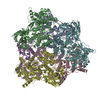
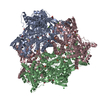
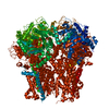
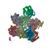
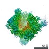
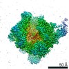
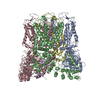
- PDB-1hcy: CRYSTAL STRUCTURE OF HEXAMERIC HAEMOCYANIN FROM PANULIRUS INTERRU... -
+ Open data

- Basic information
| Entry | Database: PDB / ID: 1hcy | |||||||||
|---|---|---|---|---|---|---|---|---|---|---|
| Title | CRYSTAL STRUCTURE OF HEXAMERIC HAEMOCYANIN FROM PANULIRUS INTERRUPTUS REFINED AT 3.2 ANGSTROMS RESOLUTION | |||||||||
 Components Components | ARTHROPODAN HEMOCYANIN | |||||||||
 Keywords Keywords |  OXYGEN TRANSPORT OXYGEN TRANSPORT | |||||||||
| Function / homology |  Function and homology informationoxygen carrier activity / oxidoreductase activity / extracellular region / metal ion binding Function and homology informationoxygen carrier activity / oxidoreductase activity / extracellular region / metal ion bindingSimilarity search - Function | |||||||||
| Biological species |  Panulirus interruptus (California spiny lobster) Panulirus interruptus (California spiny lobster) | |||||||||
| Method | X-RAY DIFFRACTION / Resolution: 3.2 Å | |||||||||
 Authors Authors | Volbeda, A. / Hol, W.G.J. | |||||||||
 Citation Citation | Journal: J.Mol.Biol. / Year: 1989 Title: Crystal structure of hexameric haemocyanin from Panulirus interruptus refined at 3.2 A resolution. Authors: Volbeda, A. / Hol, W.G. #1: Journal: Eur.J.Biochem. / Year: 1989Title: Spectroscopic Investigations of Panulirus Interruptus Hemocyanin in the Crystalline State Authors: Volbeda, A. / Feiters, M.C. / Vincent, M.G. / Bouwman, E. / Dobson, B. / Kalk, K.H. / Reedijk, J. / Hol, W.G.J. #2: Journal: J.Mol.Biol. / Year: 1989Title: Pseudo 2-Fold Symmetry in the Copper-Binding Domain of Arthropodan Haemocyanins. Possible Implications for the Evolution of Oxygen Transport Proteins Authors: Volbeda, A. / Hol, W.G.J. #3: Journal: Eur.J.Biochem. / Year: 1988Title: Panulirus Interruptus Hemocyanin. The Amino Acid Sequence of Subunit B and Anomalous Behavior of Subunits a and B on Polyacrylamide Gel Electrophoresis in the Presence of /Sds Authors: Jekel, P.A. / Bak, H.J. / Soeter, N.M. / Vereijken, J.M. / Beintema, J.J. #4: Journal: Eur.J.Biochem. / Year: 1987Title: Panulirus Interruptus Hemocyanin. The Elucidation of the Complete Amino Acid Sequence of Subunit A Authors: Bak, H.J. / Beintema, J.J. #5: Journal: J.Mol.Biol. / Year: 1986Title: Structure Determination of Panulirus Interruptus Haemocyanin at 3.2 Angstroms Resolution. Successful Phase Extension by Sixfold Density Averaging Authors: Gaykema, W.P.J. / Volbeda, A. / Hol, W.G.J. #6: Journal: Science / Year: 1985Title: The Structure of Arthropod Hemocyanins Authors: Linzen, B. / Soeter, N.M. / Riggs, A.F. / Schneider, H.-J. / Schartau, W. / Moore, M.D. / Yokota, E. / Behrens, P.Q. / Nakashima, H. / Takagi, T. / Nemoto, T. / Vereijken, J.M. / Bak, H.J. / ...Authors: Linzen, B. / Soeter, N.M. / Riggs, A.F. / Schneider, H.-J. / Schartau, W. / Moore, M.D. / Yokota, E. / Behrens, P.Q. / Nakashima, H. / Takagi, T. / Nemoto, T. / Vereijken, J.M. / Bak, H.J. / Beintema, J.J. / Volbeda, A. / Gaykema, W.P.J. / Hol, W.G.J. #7: Journal: Nature / Year: 1984Title: 3.2 Angstroms Structure of the Copper-Containing, Oxygen-Carrying Protein Panulirus Interruptus Haemocyanin Authors: Gaykema, W.P.J. / Hol, W.G.J. / Vereijken, J.M. / Soeter, N.M. / Bak, H.J. / Beintema, J.J. | |||||||||
| History |
|
- Structure visualization
| Structure viewer | Molecule: MolmilJmol/JSmol |
|---|
- Downloads & links
-Download
| PDBx/mmCIF format | 1hcy.cif.gz | 834 KB | Display | PDBx/mmCIF format |
|---|---|---|---|---|
| PDB format | pdb1hcy.ent.gz | 621.3 KB | Display | PDB format |
| PDBx/mmJSON format | 1hcy.json.gz | Tree view | PDBx/mmJSON format | |
| Others |  Other downloads Other downloads |
-Validation report
| Arichive directory | https://data.pdbj.org/pub/pdb/validation_reports/hc/1hcyftp://data.pdbj.org/pub/pdb/validation_reports/hc/1hcy | HTTPS FTP |
|---|
-Related structure data


-Links
 PDBj
PDBj- Assembly
| Deposited unit | 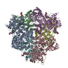
| ||||||||||||||||||||||||
|---|---|---|---|---|---|---|---|---|---|---|---|---|---|---|---|---|---|---|---|---|---|---|---|---|---|
| 1 |
| ||||||||||||||||||||||||
| Unit cell |
| ||||||||||||||||||||||||
| Atom site foot note | 1: RESIDUE PRO 625 IS A CIS PROLINE. | ||||||||||||||||||||||||
| Noncrystallographic symmetry (NCS) | NCS oper:
| ||||||||||||||||||||||||
| Details | THE TRANSFORMATIONS PRESENTED ON *MTRIX* RECORDS BELOW CAN BE USED TO GENERATE THE COMPLETE HEXAMER. |
-Components
| #1: Protein | Mass: 75696.203 Da / Num. of mol.: 6 / Source method: isolated from a natural source Source: (natural) Panulirus interruptus (California spiny lobster)References: UniProt: P04254 #2: Polysaccharide | 2-acetamido-2-deoxy-beta-D-glucopyranose-(1-4)-2-acetamido-2-deoxy-beta-D-glucopyranose / Mass: 424.401 Da / Num. of mol.: 6Source method: isolated from a genetically manipulated source #3: Chemical | Copper  Mass: 63.546 Da / Num. of mol.: 2 / Source method: obtained synthetically / Formula: Cu Mass: 63.546 Da / Num. of mol.: 2 / Source method: obtained synthetically / Formula: Cu#4: Water | ChemComp-HOH / | Water Mass: 18.015 Da / Num. of mol.: 186 / Source method: isolated from a natural source / Formula: H2O Mass: 18.015 Da / Num. of mol.: 186 / Source method: isolated from a natural source / Formula: H2OCompound details | PANULIRUS INTERRUPTUS HAEMOCYANIN HEXAMERS CONTAIN THREE DIFFERENT, THOUGH HOMOLOGOUS, SUBUNIT ...PANULIRUS INTERRUPTU | |
|---|
-Experimental details
-Experiment
| Experiment | Method: X-RAY DIFFRACTION |
|---|
- Sample preparation
| Crystal | Density Matthews: 2.74 Å3/Da / Density % sol: 55.18 % | |||||||||||||||
|---|---|---|---|---|---|---|---|---|---|---|---|---|---|---|---|---|
| Crystal grow | *PLUS Temperature: 4 ℃ / Method: microdialysis / Details: referred to J.Mol.Biol. 99.619-629 / PH range low: 4.2 / PH range high: 4 | |||||||||||||||
| Components of the solutions | *PLUS
|
-Data collection
| Radiation | Scattering type: x-ray |
|---|---|
| Radiation wavelength | Relative weight: 1 |
- Processing
| Software |
| |||||||||||||||||||||||||||||||||||||||||||||||||||||||||||||||
|---|---|---|---|---|---|---|---|---|---|---|---|---|---|---|---|---|---|---|---|---|---|---|---|---|---|---|---|---|---|---|---|---|---|---|---|---|---|---|---|---|---|---|---|---|---|---|---|---|---|---|---|---|---|---|---|---|---|---|---|---|---|---|---|---|
| Refinement | Resolution: 3.2→8 Å Details: RESIDUES 1 - 4, 171 - 174, 551 - 552, 658 - 659 (THE TWO N-ACETYL GLUCOSAMINES) AND 34 WATER MOLECULES WITH TEMPERATURE FACTORS ABOVE 50 SHOULD BE CONSIDERED UNRELIABLE. IF THESE RESIDUES ...Details: RESIDUES 1 - 4, 171 - 174, 551 - 552, 658 - 659 (THE TWO N-ACETYL GLUCOSAMINES) AND 34 WATER MOLECULES WITH TEMPERATURE FACTORS ABOVE 50 SHOULD BE CONSIDERED UNRELIABLE. IF THESE RESIDUES AND WATERS ARE EXCLUDED FROM THE REFINEMENT, THE R-FACTOR BECOMES 0.224. THE REFINEMENT WAS CARRIED OUT BY FIRST MAINTAINING PERFECT 32 SYMMETRY, FOLLOWED BY RIGID BODY REFINEMENT OF THE 6 SUB UNITS, RESULTING IN A HEXAMER OF IDENTICAL SUB UNITS, BUT WITH SIGNIFICANT DEVIATIONS FROM 32 SYMMETRY (PROTEIN DATA BANK ENTRY 1HCY). SUBSEQUENT REFINEMENT RELAXING THE LOCAL SYMMETRY CONSTRAINTS RESULTED IN SIX NON-IDENTICAL MODELS (PROTEIN DATA BANK ENTRIES 1HC1 - 1HC6). NOTE THAT THE FOLLOWING DISTANCES ARE UNUSUALLY LARGE: C7 NAG 658 - C8 NAG 658 2.884 ANGSTROMS C7 NAG 659 - C8 NAG 659 3.140 ANGSTROMS
| |||||||||||||||||||||||||||||||||||||||||||||||||||||||||||||||
| Refinement step | Cycle: LAST / Resolution: 3.2→8 Å
| |||||||||||||||||||||||||||||||||||||||||||||||||||||||||||||||
| Refine LS restraints |
| |||||||||||||||||||||||||||||||||||||||||||||||||||||||||||||||
| Refinement | *PLUS Highest resolution: 3.2 Å / Lowest resolution: 8 Å / Num. reflection obs: 59193 / Rfactor obs: 0.221 | |||||||||||||||||||||||||||||||||||||||||||||||||||||||||||||||
| Solvent computation | *PLUS | |||||||||||||||||||||||||||||||||||||||||||||||||||||||||||||||
| Displacement parameters | *PLUS | |||||||||||||||||||||||||||||||||||||||||||||||||||||||||||||||
| Refine LS restraints | *PLUS
|
