Group: Data collection / Category: diffrn_source / Item: _diffrn_source.pdbx_synchrotron_site
Remark 700
SHEET DETERMINATION METHOD: DSSP THE SHEETS PRESENTED AS "A" IN EACH CHAIN ON SHEET RECORDS BELOW ... SHEET DETERMINATION METHOD: DSSP THE SHEETS PRESENTED AS "A" IN EACH CHAIN ON SHEET RECORDS BELOW IS ACTUALLY AN 8-STRANDED BARREL THIS IS REPRESENTED BY A 9-STRANDED SHEET IN WHICH THE FIRST AND LAST STRANDS ARE IDENTICAL.
Mass: 18.015 Da / Num. of mol.: 538 / Source method: isolated from a natural source / Formula: H2O
Compound details
STRUCTURE WAS SOLVED IN THE ABSENCE OF CHEMICAL SEQUENCE. PROTECT CELLS FROM THE TOXIC EFFECTS OF ...STRUCTURE WAS SOLVED IN THE ABSENCE OF CHEMICAL SEQUENCE. PROTECT CELLS FROM THE TOXIC EFFECTS OF HYDROGEN PEROXIDE BY BREAKING DOWN HYDROGEN PEROXIDE IN WATER AND OXYGEN.
-
Experimental details
-
Experiment
Experiment
Method: X-RAY DIFFRACTION / Number of used crystals: 1
-
Sample preparation
Crystal
Density Matthews: 2.69 Å3/Da / Density % sol: 54.2 %
Protocol: SINGLE WAVELENGTH / Monochromatic (M) / Laue (L): M / Scattering type: x-ray
Radiation wavelength
Wavelength: 0.98 Å / Relative weight: 1
Reflection
Resolution: 1.501→12 Å / Num. obs: 358628 / % possible obs: 97 % / Redundancy: 3.7 % / Rmerge(I) obs: 0.06 / Net I/σ(I): 7.9
Reflection shell
Resolution: 1.5→1.54 Å / Redundancy: 3.4 % / Rmerge(I) obs: 0.21 / Mean I/σ(I) obs: 3.6 / % possible all: 96
Reflection
*PLUS
Num. obs: 95355 / Num. measured all: 358628
-
Processing
Software
Name
Version
Classification
REFMAC
5.0.36
refinement
MOSFLM
datascaling
Refinement
Method to determine structure: MIR / Resolution: 1.5→12 Å / Cor.coef. Fo:Fc: 0.988 / Cor.coef. Fo:Fc free: 0.981 / SU B: 0.59 / SU ML: 0.022 / Cross valid method: THROUGHOUT / ESU R: 0.043 / ESU R Free: 0.042 / Stereochemistry target values: MAXIMUM LIKELIHOOD / Details: HYDROGENS HAVE BEEN ADDED IN THE RIDING POSITIONS
Rfactor
Num. reflection
% reflection
Selection details
Rfree
0.12
4781
5 %
RANDOM
Rwork
0.092
-
-
-
obs
-
90574
97.5 %
-
Solvent computation
Ion probe radii: 0.8 Å / Shrinkage radii: 0.8 Å / VDW probe radii: 1.4 Å / Solvent model: BABINET MODEL PLUS MASK
 Movie
Movie Controller


 Open data
 Basic information
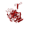
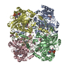
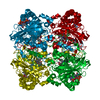
 Components
 Keywords
 Function and homology information
 Authors
 Citation
 Structure visualization
 Downloads & links
 Other downloads
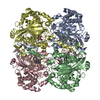
 PDBj
PDBj
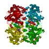
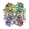
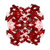
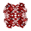
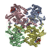
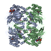
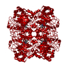
 Assembly
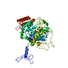

 Mass: 616.487 Da / Num. of mol.: 1 / Source method: obtained synthetically / Formula: C34H32FeN4O4
 Mass: 96.063 Da / Num. of mol.: 1 / Source method: obtained synthetically / Formula: SO4
 Mass: 18.015 Da / Num. of mol.: 538 / Source method: isolated from a natural source / Formula: H2O
 Sample preparation
 / Beamline: X31 / Wavelength: 0.98
  Processing