 Movie
Movie Controller

[English] 日本語
 Yorodumi
- PDB-1hbt: Human alpha-thrombin complexed with a peptidyl pyridinium methyl ... -
+ Open data

- Basic information
| Entry | Database: PDB / ID: 1hbt | |||||||||
|---|---|---|---|---|---|---|---|---|---|---|
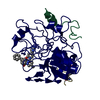
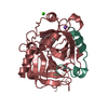
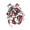
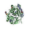
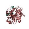
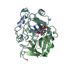
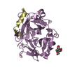
| Title | Human alpha-thrombin complexed with a peptidyl pyridinium methyl ketone containing bivalent inhibitor | |||||||||
 Components Components |
| |||||||||
 Keywords Keywords | HYDROLASE/HYDROLASE INHIBITOR /  SERINE PROTEASE / HYDROLASE-HYDROLASE INHIBITOR COMPLEX SERINE PROTEASE / HYDROLASE-HYDROLASE INHIBITOR COMPLEX | |||||||||
| Function / homology |  Function and homology information Function and homology informationpositive regulation of lipid kinase activity / positive regulation of phospholipase C-activating G protein-coupled receptor signaling pathway / cytolysis by host of symbiont cells / thrombospondin receptor activity / Defective factor XII causes hereditary angioedema / thrombin / neutrophil-mediated killing of gram-negative bacterium / regulation of blood coagulation / ligand-gated ion channel signaling pathway / Defective F8 cleavage by thrombin ...positive regulation of lipid kinase activity / positive regulation of phospholipase C-activating G protein-coupled receptor signaling pathway / cytolysis by host of symbiont cells / thrombospondin receptor activity / Defective factor XII causes hereditary angioedema / thrombin / neutrophil-mediated killing of gram-negative bacterium / regulation of blood coagulation / ligand-gated ion channel signaling pathway / Defective F8 cleavage by thrombin / Platelet Aggregation (Plug Formation) / negative regulation of platelet activation / negative regulation of astrocyte differentiation / positive regulation of collagen biosynthetic process / negative regulation of cytokine production involved in inflammatory response / positive regulation of blood coagulation / negative regulation of fibrinolysis / Gamma-carboxylation of protein precursors / Transport of gamma-carboxylated protein precursors from the endoplasmic reticulum to the Golgi apparatus / Common Pathway of Fibrin Clot Formation / Removal of aminoterminal propeptides from gamma-carboxylated proteins / fibrinolysis / regulation of cytosolic calcium ion concentration / Intrinsic Pathway of Fibrin Clot Formation / Peptide ligand-binding receptors / positive regulation of release of sequestered calcium ion into cytosol / Regulation of Complement cascade / acute-phase response / Cell surface interactions at the vascular wall / lipopolysaccharide binding / negative regulation of proteolysis / positive regulation of receptor signaling pathway via JAK-STAT / growth factor activity / positive regulation of insulin secretion / platelet activation / response to wounding / Golgi lumen / positive regulation of protein localization to nucleus / Regulation of Insulin-like Growth Factor (IGF) transport and uptake by Insulin-like Growth Factor Binding Proteins (IGFBPs) / positive regulation of reactive oxygen species metabolic process / blood coagulation / antimicrobial humoral immune response mediated by antimicrobial peptide / Thrombin signalling through proteinase activated receptors (PARs) / heparin binding / regulation of cell shape / positive regulation of cell growth / G alpha (q) signalling events / collagen-containing extracellular matrix / blood microparticle / cell surface receptor signaling pathway / positive regulation of phosphatidylinositol 3-kinase/protein kinase B signal transduction / positive regulation of protein phosphorylation / G protein-coupled receptor signaling pathway / endoplasmic reticulum lumen / signaling receptor binding / serine-type endopeptidase activity / calcium ion binding / positive regulation of cell population proliferation / proteolysis / extracellular space / extracellular exosome / extracellular region / plasma membraneSimilarity search - Function | |||||||||
| Biological species |  Homo sapiens (human) Homo sapiens (human) | |||||||||
| Method | X-RAY DIFFRACTION / Resolution: 2 Å | |||||||||
 Authors Authors | Rehse, P.H. / Cygler, M. | |||||||||
 Citation Citation | Journal: Biochemistry / Year: 1995 Title: Crystal structure of a peptidyl pyridinium methyl ketone inhibitor with thrombin. Authors: Rehse, P.H. / Steinmetzer, T. / Li, Y. / Konishi, Y. / Cygler, M. | |||||||||
| History |
|
- Structure visualization
| Structure viewer | Molecule: MolmilJmol/JSmol |
|---|
- Downloads & links
-Download
| PDBx/mmCIF format | 1hbt.cif.gz | 80 KB | Display | PDBx/mmCIF format |
|---|---|---|---|---|
| PDB format | pdb1hbt.ent.gz | 57.3 KB | Display | PDB format |
| PDBx/mmJSON format | 1hbt.json.gz | Tree view | PDBx/mmJSON format | |
| Others |  Other downloads Other downloads |
-Validation report
| Arichive directory | https://data.pdbj.org/pub/pdb/validation_reports/hb/1hbtftp://data.pdbj.org/pub/pdb/validation_reports/hb/1hbt | HTTPS FTP |
|---|
-Related structure data
| Similar structure data |
|---|


-Links
 PDBj
PDBj


- Assembly
| Deposited unit | 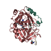
| |||||||||
|---|---|---|---|---|---|---|---|---|---|---|
| 1 |
| |||||||||
| Unit cell |
| |||||||||
| Atom site foot note | 1: CIS PROLINE - PRO H 37 | |||||||||
| Components on special symmetry positions |
|

-Components
| #1: Protein/peptide | Mass: 4096.534 Da / Num. of mol.: 1 Source method: isolated from a genetically manipulated source Source: (gene. exp.) Homo sapiens (human) / Gene: DKFZp470K2111, F2 / References: UniProt: P00734, thrombin |
|---|---|
| #2: Protein | Mass: 29780.219 Da / Num. of mol.: 1 Source method: isolated from a genetically manipulated source Source: (gene. exp.) Homo sapiens (human) / Gene: F2 / References: UniProt: P00734, thrombin |
| #3: Protein/peptide | Mass: 2116.328 Da / Num. of mol.: 1 / Source method: obtained synthetically |
| #4: Water | ChemComp-HOH / Water Mass: 18.015 Da / Num. of mol.: 185 / Source method: isolated from a natural source / Formula: H2O Mass: 18.015 Da / Num. of mol.: 185 / Source method: isolated from a natural source / Formula: H2O |
| Compound details | THE FORMATION OF HEMIKETAL AT POSITION C2 OF AR0 RESIDUE IS A RESULT OF NUCLEOPHILIC ATTACK OF OG ...THE FORMATION OF HEMIKETAL AT POSITION C2 OF AR0 RESIDUE IS A RESULT OF NUCLEOPHIL |
| Sequence details | THROMBIN IS CLEAVED BETWEEN RESIDUES 15 AND 16. CHAIN ID L IS USED FOR RESIDUES 1 - 15 AND CHAIN ID ...THROMBIN IS CLEAVED BETWEEN RESIDUES 15 AND 16. CHAIN ID L IS USED FOR RESIDUES 1 - 15 AND CHAIN ID H IS USED FOR RESIDUES 16 - 247. CHAIN ID I IS USED FOR THE INHIBITOR P596 |
-Experimental details
-Experiment
| Experiment | Method: X-RAY DIFFRACTION |
|---|
- Sample preparation
| Crystal | Density Matthews: 2.54 Å3/Da / Density % sol: 51.48 % | ||||||||||||||||||
|---|---|---|---|---|---|---|---|---|---|---|---|---|---|---|---|---|---|---|---|
| Crystal grow | *PLUS Temperature: 18 ℃ / pH: 6.2 / Method: vapor diffusion, hanging drop | ||||||||||||||||||
| Components of the solutions | *PLUS
|
-Data collection
| Detector | Date: Aug 16, 1994 |
|---|---|
| Radiation | Monochromatic (M) / Laue (L): M / Scattering type: x-ray |
| Radiation wavelength | Relative weight: 1 |
| Reflection | Resolution: 2→20 Å / Num. obs: 19998 / % possible obs: 82.1 % / Observed criterion σ(I): 1 / Redundancy: 2.2 % / Rmerge(I) obs: 0.082 |
| Reflection | *PLUS Num. measured all: 43266 / Rmerge(I) obs: 0.082 |
| Reflection shell | *PLUS Highest resolution: 2 Å / Lowest resolution: 2.1 Å / % possible obs: 69 % |
- Processing
| Software |
| ||||||||||||||||||||||||||||||||||||||||||||||||||||||||||||
|---|---|---|---|---|---|---|---|---|---|---|---|---|---|---|---|---|---|---|---|---|---|---|---|---|---|---|---|---|---|---|---|---|---|---|---|---|---|---|---|---|---|---|---|---|---|---|---|---|---|---|---|---|---|---|---|---|---|---|---|---|---|
| Refinement | Resolution: 2→8 Å / σ(F): 3 Details: RESIDUES I 51 - I 54 WERE FITTED TO AN OMIT MAP. THE REGION IS HIGHLY FLEXIBLE AND POSSIBLY ADOPTS MULTIPLE CONFORMATIONS. THE ENTIRE SIDE CHAIN OF RESIDUE I 61 WAS NOT OBSERVABLE AND WAS ...Details: RESIDUES I 51 - I 54 WERE FITTED TO AN OMIT MAP. THE REGION IS HIGHLY FLEXIBLE AND POSSIBLY ADOPTS MULTIPLE CONFORMATIONS. THE ENTIRE SIDE CHAIN OF RESIDUE I 61 WAS NOT OBSERVABLE AND WAS NOT MODELED. THE COVALENT BOND FORMED BETWEEN THE SER H 195 OG OF THROMBIN AND THE C2 ATOM OF THE AR0 I 3 CAUSES THE LATTER ATOM TO BECOME TETRAHEDRAL. WATER 1184 AND THE SIDE CHAIN BEYOND CB ARG H 75 WERE EXCLUDED SINCE THEY OCCUPIED NEARLY THE SAME SPACE ALONG A TWO-FOLD. IN THE ENTRY THEY ARE GIVEN AN OCCUPANCY OF 0.5. ALTERNATE CONFORMATION OF THE ARG H 75 SIDE CHAIN WAS NOT OBVIOUS.
| ||||||||||||||||||||||||||||||||||||||||||||||||||||||||||||
| Displacement parameters | Biso mean: 27.97 Å2 | ||||||||||||||||||||||||||||||||||||||||||||||||||||||||||||
| Refinement step | Cycle: LAST / Resolution: 2→8 Å
| ||||||||||||||||||||||||||||||||||||||||||||||||||||||||||||
| Refine LS restraints |
| ||||||||||||||||||||||||||||||||||||||||||||||||||||||||||||
| Software | *PLUS Name: X-PLOR / Classification: refinement | ||||||||||||||||||||||||||||||||||||||||||||||||||||||||||||
| Refinement | *PLUS Rfactor all: 0.21 | ||||||||||||||||||||||||||||||||||||||||||||||||||||||||||||
| Solvent computation | *PLUS | ||||||||||||||||||||||||||||||||||||||||||||||||||||||||||||
| Displacement parameters | *PLUS | ||||||||||||||||||||||||||||||||||||||||||||||||||||||||||||
| Refine LS restraints | *PLUS
|
