 Movie
Movie Controller

[English] 日本語
 Yorodumi
- PDB-1h8h: Bovine mitochondrial F1-ATPase crystallised in the presence of 5m... -
+ Open data

- Basic information
| Entry | Database: PDB / ID: 1h8h | ||||||
|---|---|---|---|---|---|---|---|
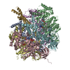
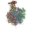
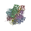
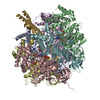
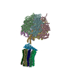
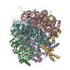
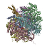
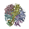
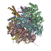
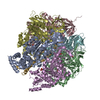
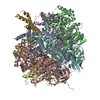
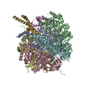
| Title | Bovine mitochondrial F1-ATPase crystallised in the presence of 5mm AMPPNP | ||||||
 Components Components | (BOVINE MITOCHONDRIAL F1- ...) x 3 | ||||||
 Keywords Keywords |  HYDROLASE / ATP PHOSPHORYLASE / ATP PHOSPHORYLASE (H+ TRANSPORTING) / ATP SYNTHASE / F1FO ATP SYNTHASE / F1-ATPASE HYDROLASE / ATP PHOSPHORYLASE / ATP PHOSPHORYLASE (H+ TRANSPORTING) / ATP SYNTHASE / F1FO ATP SYNTHASE / F1-ATPASE | ||||||
| Function / homology |  Function and homology information Function and homology informationFormation of ATP by chemiosmotic coupling / Cristae formation / mitochondrial proton-transporting ATP synthase, catalytic core / mitochondrial proton-transporting ATP synthase complex / mitochondrial proton-transporting ATP synthase complex, catalytic sector F(1) / proton motive force-driven mitochondrial ATP synthesis / proton motive force-driven ATP synthesis / proton-transporting ATP synthase complex, catalytic core F(1) / H+-transporting two-sector ATPase / proton-transporting ATPase activity, rotational mechanism ...Formation of ATP by chemiosmotic coupling / Cristae formation / mitochondrial proton-transporting ATP synthase, catalytic core / mitochondrial proton-transporting ATP synthase complex / mitochondrial proton-transporting ATP synthase complex, catalytic sector F(1) / proton motive force-driven mitochondrial ATP synthesis / proton motive force-driven ATP synthesis / proton-transporting ATP synthase complex, catalytic core F(1) / H+-transporting two-sector ATPase / proton-transporting ATPase activity, rotational mechanism / proton-transporting ATP synthase activity, rotational mechanism / ADP binding / ATP hydrolysis activity / ATP binding / plasma membraneSimilarity search - Function | ||||||
| Biological species |  BOS TAURUS (cattle) BOS TAURUS (cattle) | ||||||
| Method | X-RAY DIFFRACTION / SYNCHROTRON / MOLECULAR REPLACEMENT / Resolution: 2.9 Å | ||||||
 Authors Authors | Braig, K. / Menz, R.I. / Montgomery, M.G. / Leslie, A.G.W. / Walker, J.E. | ||||||
 Citation Citation | Journal: FEBS Lett. / Year: 2001 Title: The Structure and Nucleotide Occupancy of Bovine Mitochondrial F(1)-ATPase are not Influenced by Crystallisation at High Concentrations of Nucleotide Authors: Menz, R.I. / Leslie, A.G.W. / Walker, J.E. #1: Journal: Nature / Year: 1994Title: Structure at 2.8 A Resolution of F1-ATPase from Bovine Heart Mitochondria Authors: Abrahams, J.P. / Leslie, A.G.W. / Lutter, R. / Walker, J.E. #2: Journal: J.Mol.Biol. / Year: 1993 Title: Crystallization of F1-ATPase from Bovine Heart Mitochondria Authors: Lutter, R. / Abrahams, J.P. / Van Raaij, M.J. / Todd, R.J. / Lundqvist, T. / Buchanan, S.K. / Leslie, A.G. / Walker, J.E. #3: Journal: Embo J. / Year: 1993 Title: Inherent Asymmetry of the Structure of F1-ATPase from Bovine Heart Mitochondria at 6.5 A Resolution Authors: Abrahams, J.P. / Lutter, R. / Todd, R.J. / Van Raaij, M.J. / Leslie, A.G. / Walker, J.E. | ||||||
| History |
|
- Structure visualization
| Structure viewer | Molecule: MolmilJmol/JSmol |
|---|
- Downloads & links
-Download
| PDBx/mmCIF format | 1h8h.cif.gz | 572.7 KB | Display | PDBx/mmCIF format |
|---|---|---|---|---|
| PDB format | pdb1h8h.ent.gz | 463.9 KB | Display | PDB format |
| PDBx/mmJSON format | 1h8h.json.gz | Tree view | PDBx/mmJSON format | |
| Others |  Other downloads Other downloads |
-Validation report
| Arichive directory | https://data.pdbj.org/pub/pdb/validation_reports/h8/1h8hftp://data.pdbj.org/pub/pdb/validation_reports/h8/1h8h | HTTPS FTP |
|---|
-Related structure data


-Links
 PDBj
PDBj


- Assembly
| Deposited unit | 
| ||||||||
|---|---|---|---|---|---|---|---|---|---|
| 1 |
| ||||||||
| Unit cell |
| ||||||||
| Details | THE F1-ATPASE MOLECULE HAS THREE COPIES OF THE NON-CATALYTICALPHA SUBUNIT AND THREE COPIES OF THE CATALYTIC BETA SUBUNIT.IN THE 1994 REFERENCE, THE BETA SUBUNITS WERE LABELED ACCORDINGTO THE BOUND NUCLEOTIDE, ASBETA(DP) (BINDS ADP),BETA(E) (NO BOUND NUCLEOTIDE) ANDBETA(TP ) (AMPPNP BOUND).THE ALPHA SUBUNITS (WHICH ALL BIND AMPPNP) CONTRIBUTE TO THECATALYTIC SITES OF THE BETA SUBUNITS, AND HAVE BEEN LABELEDACCORDINGLY . THUSALPHA(DP) CONTRIBUTES TO THE CATALYTIC SITE ON BETA(DP),ALPHA(TP) TO THE SITE ON BETA ( TP) ANDALPHA(E) TO THE SITE ON BETA(E).THE CORRESPONDENCE BETWEEN THE SUBUNIT NAMES AND THE CHAINIDENTIFIERS IS GIVEN BELOW:.CHAIN A: ALPHA(E )CHAIN B: ALPHA(TP)CHAIN C: ALPHA(DP)CHAIN D : BETA(DP)CHAIN E: BETA(E)CHAIN F: BETA(TP) CHAIN G: GAMMA SUBUNIT |
-Components
-BOVINE MITOCHONDRIAL F1- ... , 3 types, 7 molecules ABCDEFG
| #1: Protein | Mass: 55301.207 Da / Num. of mol.: 3 / Source method: isolated from a natural source / Source: (natural) BOS TAURUS (cattle) / Organ: HEART / Organelle: MITOCHONDRION / Tissue: MUSCLESkeletal muscle / References: UniProt: P19483, EC: 3.6.1.34#2: Protein | Mass: 51757.836 Da / Num. of mol.: 3 / Source method: isolated from a natural source / Source: (natural) BOS TAURUS (cattle) / Organ: HEART / Organelle: MITOCHONDRION / Tissue: MUSCLESkeletal muscle / References: UniProt: P00829, EC: 3.6.1.34#3: Protein | | Mass: 30185.674 Da / Num. of mol.: 1 / Source method: isolated from a natural source / Source: (natural) BOS TAURUS (cattle) / Organ: HEART / Organelle: MITOCHONDRION / Tissue: MUSCLESkeletal muscle / References: UniProt: P05631, EC: 3.6.1.34 |
|---|
-Non-polymers , 6 types, 123 molecules 


| #4: Chemical | ChemComp-ATP / Adenosine triphosphate Mass: 507.181 Da / Num. of mol.: 4 / Source method: obtained synthetically / Formula: C10H16N5O13P3 / Comment: ATP, energy-carrying molecule*YM Mass: 507.181 Da / Num. of mol.: 4 / Source method: obtained synthetically / Formula: C10H16N5O13P3 / Comment: ATP, energy-carrying molecule*YM#5: Chemical | ChemComp-MG /  Mass: 24.305 Da / Num. of mol.: 5 / Source method: obtained synthetically / Formula: Mg Mass: 24.305 Da / Num. of mol.: 5 / Source method: obtained synthetically / Formula: Mg#6: Chemical | ChemComp-GOL / | Glycerol Mass: 92.094 Da / Num. of mol.: 1 / Source method: obtained synthetically / Formula: C3H8O3 Mass: 92.094 Da / Num. of mol.: 1 / Source method: obtained synthetically / Formula: C3H8O3#7: Chemical | ChemComp-ADP / | Adenosine diphosphate Mass: 427.201 Da / Num. of mol.: 1 / Source method: obtained synthetically / Formula: C10H15N5O10P2 / Comment: ADP, energy-carrying molecule*YM Mass: 427.201 Da / Num. of mol.: 1 / Source method: obtained synthetically / Formula: C10H15N5O10P2 / Comment: ADP, energy-carrying molecule*YM#8: Chemical | ChemComp-PO4 / | Phosphate Mass: 94.971 Da / Num. of mol.: 1 / Source method: obtained synthetically / Formula: PO4 Mass: 94.971 Da / Num. of mol.: 1 / Source method: obtained synthetically / Formula: PO4#9: Water | ChemComp-HOH / | WaterMass: 18.015 Da / Num. of mol.: 111 / Source method: isolated from a natural source / Formula: H2O |
|---|
-Details
| Sequence details | REFERENCE: 1) FOR THE ALPHA SUBUNIT: J. E. WALKER, S. J. POWELL, O. VINAS AND M. J. RUNSWICK, ...REFERENCE: 1) FOR THE ALPHA SUBUNIT: J. E. WALKER, S. J. POWELL, O. VINAS AND M. J. RUNSWICK, BIOCHEMIST |
|---|
-Experimental details
-Experiment
| Experiment | Method: X-RAY DIFFRACTION / Number of used crystals: 1 |
|---|
- Sample preparation
| Crystal | Density Matthews: 2.97 Å3/Da / Density % sol: 54 % | ||||||||||||||||||||||||||||||||||||||||||||||||||||||||||||||||||||||||||||||||
|---|---|---|---|---|---|---|---|---|---|---|---|---|---|---|---|---|---|---|---|---|---|---|---|---|---|---|---|---|---|---|---|---|---|---|---|---|---|---|---|---|---|---|---|---|---|---|---|---|---|---|---|---|---|---|---|---|---|---|---|---|---|---|---|---|---|---|---|---|---|---|---|---|---|---|---|---|---|---|---|---|---|
| Crystal grow | pH: 8 Details: CRYSTALS WERE GROWN IN THE PRESENCE OF AZIDE, A KNOWN INHIBITOR, BUT THIS HAS NOT BEEN LOCATED IN THE STRUCTURE., pH 8.00 | ||||||||||||||||||||||||||||||||||||||||||||||||||||||||||||||||||||||||||||||||
| Crystal grow | *PLUS Method: microdialysisDetails: solution 2 also includes 0.02%(w/v) sodium azide, 0.001%(w/v) phenylmethylsulphonyl fluoride, 5 mM dithiothreitol, 5mM AMPPNP, 0.005mM ADP and 11-12%(w/v) PEG6000 | ||||||||||||||||||||||||||||||||||||||||||||||||||||||||||||||||||||||||||||||||
| Components of the solutions | *PLUS
|
-Data collection
| Diffraction | Mean temperature: 100 K |
|---|---|
| Diffraction source | Source: SYNCHROTRON / Site: ESRF  / Beamline: BM14 / Wavelength: 0.953 / Beamline: BM14 / Wavelength: 0.953 |
| Detector | Type: MARRESEARCH / Detector: IMAGE PLATE / Date: Sep 18, 1997 |
| Radiation | Protocol: SINGLE WAVELENGTH / Monochromatic (M) / Laue (L): M / Scattering type: x-ray |
| Radiation wavelength | Wavelength: 0.953 Å / Relative weight: 1 |
| Reflection | Resolution: 2.88→37.27 Å / Num. obs: 90077 / % possible obs: 95.8 % / Observed criterion σ(I): 0 / Redundancy: 2.5 % / Biso Wilson estimate: 62.2 Å2 / Rmerge(I) obs: 0.063 / Net I/σ(I): 13.5 |
| Reflection shell | Resolution: 2.87→3.07 Å / Redundancy: 2.3 % / Rmerge(I) obs: 0.166 / Mean I/σ(I) obs: 7.3 / % possible all: 90.4 |
| Reflection | *PLUS Num. obs: 79330 / Num. measured all: 217558 |
| Reflection shell | *PLUS % possible obs: 90.4 % |
- Processing
| Software |
| ||||||||||||||||||||||||||||||||||||||||||||||||||||||||||||||||||||||||||||||||||||
|---|---|---|---|---|---|---|---|---|---|---|---|---|---|---|---|---|---|---|---|---|---|---|---|---|---|---|---|---|---|---|---|---|---|---|---|---|---|---|---|---|---|---|---|---|---|---|---|---|---|---|---|---|---|---|---|---|---|---|---|---|---|---|---|---|---|---|---|---|---|---|---|---|---|---|---|---|---|---|---|---|---|---|---|---|---|
| Refinement | Method to determine structure: MOLECULAR REPLACEMENT Starting model: PDB CODE 1E1Q, BOVINE MITOCHONDRIAL F1-ATPASE AT 100K Resolution: 2.9→20 Å / SU B: 16.8 / SU ML: 0.34 / Cross valid method: THROUGHOUT / σ(F): 0 / ESU R Free: 0.45 Details: RESIDUES B 402 - B 409 INCLUSIVE HAVE BEEN GIVEN ZERO OCCUPANCY AS THERE WAS NO INTERPRETABLE ELECTRON DENSITY IN THIS REGION. THE POSITIONS OF SIDE CHAIN ATOMS WITH TEMPERATURE FACTORS ...Details: RESIDUES B 402 - B 409 INCLUSIVE HAVE BEEN GIVEN ZERO OCCUPANCY AS THERE WAS NO INTERPRETABLE ELECTRON DENSITY IN THIS REGION. THE POSITIONS OF SIDE CHAIN ATOMS WITH TEMPERATURE FACTORS GREATER THAN 75 IS UNCERTAIN. THE MAIN CHAIN CONFORMATION IS ALSO UNCERTAIN FOR REGIONS WITH TEMPERATURE FACTORS ABOVE 60. THE PEPTIDE BOND BETWEEN ASP 269 AND ASP 270 IN CHAINS A, B, C AND THE PEPTIDE BOND BETWEEN ASP 256 AND ASN 257 IN CHAINS D, E, AND F HAVE BEEN MODELED IN A CIS CONFORMATION.
| ||||||||||||||||||||||||||||||||||||||||||||||||||||||||||||||||||||||||||||||||||||
| Displacement parameters | Biso mean: 45.3 Å2 | ||||||||||||||||||||||||||||||||||||||||||||||||||||||||||||||||||||||||||||||||||||
| Refinement step | Cycle: LAST / Resolution: 2.9→20 Å
| ||||||||||||||||||||||||||||||||||||||||||||||||||||||||||||||||||||||||||||||||||||
| Refine LS restraints |
|
