 Movie
Movie Controller

[English] 日本語
 Yorodumi
- PDB-1h7h: The structure of CMP:2-keto-3-deoxy-manno-octonic acid synthetase... -
+ Open data

- Basic information
| Entry | Database: PDB / ID: 1h7h | ||||||
|---|---|---|---|---|---|---|---|
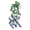
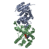
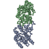
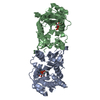
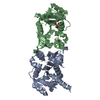
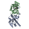
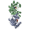
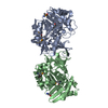
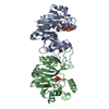
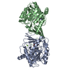
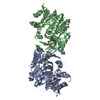
| Title | The structure of CMP:2-keto-3-deoxy-manno-octonic acid synthetase and of its complexes with substrates and substrate analogues, CDP complex | ||||||
 Components Components | 3-DEOXY-MANNO-OCTULOSONATE CYTIDYLYLTRANSFERASE | ||||||
 Keywords Keywords | NUCLEOTIDYLTRANSFERASE / CMP-KDO SYNTHETASE / NUCLEOSIDE MONOPHOSPHATE GLYCOSIDES / LIPOPOLYSACCHARIDE BIOSYNTHESIS / SUGAR-ACTIVATING ENZYMES | ||||||
| Function / homology |  Function and homology information3-deoxy-manno-octulosonate cytidylyltransferase / 3-deoxy-manno-octulosonate cytidylyltransferase activity / CMP-keto-3-deoxy-D-manno-octulosonic acid biosynthetic process / lipopolysaccharide biosynthetic process / cytoplasm Function and homology information3-deoxy-manno-octulosonate cytidylyltransferase / 3-deoxy-manno-octulosonate cytidylyltransferase activity / CMP-keto-3-deoxy-D-manno-octulosonic acid biosynthetic process / lipopolysaccharide biosynthetic process / cytoplasmSimilarity search - Function | ||||||
| Biological species |  ESCHERICHIA COLI (E. coli) ESCHERICHIA COLI (E. coli) | ||||||
| Method | X-RAY DIFFRACTION / OTHER / Resolution: 2.3 Å | ||||||
 Authors Authors | Jelakovic, S. / Schulz, G.E. | ||||||
 Citation Citation | Journal: J.Mol.Biol. / Year: 2001 Title: The Structure of Cmp:2-Keto-3-Deoxy-Manno-Octonic Acid Synthetase and of its Complexes with Substrates and Substrate Analogs Authors: Jelakovic, S. / Schulz, G.E. #1: Journal: FEBS Lett. / Year: 1996Title: The Three-Dimensional Structure of Capsule-Specific Cmp:2-Keto-3-Deoxy-Manno-Octonic Acid Synthetase from Escherichia Coli Authors: Jelakovic, S. / Jann, K. / Schulz, G.E. | ||||||
| History |
|
- Structure visualization
| Structure viewer | Molecule: MolmilJmol/JSmol |
|---|
- Downloads & links
-Download
| PDBx/mmCIF format | 1h7h.cif.gz | 108.2 KB | Display | PDBx/mmCIF format |
|---|---|---|---|---|
| PDB format | pdb1h7h.ent.gz | 88.3 KB | Display | PDB format |
| PDBx/mmJSON format | 1h7h.json.gz | Tree view | PDBx/mmJSON format | |
| Others |  Other downloads Other downloads |
-Validation report
| Arichive directory | https://data.pdbj.org/pub/pdb/validation_reports/h7/1h7hftp://data.pdbj.org/pub/pdb/validation_reports/h7/1h7h | HTTPS FTP |
|---|
-Related structure data


-Links
 PDBj
PDBj

- Assembly
| Deposited unit | 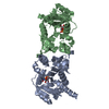
| ||||||||
|---|---|---|---|---|---|---|---|---|---|
| 1 |
| ||||||||
| Unit cell |
| ||||||||
| Noncrystallographic symmetry (NCS) | NCS oper: (Code: given Matrix: (-0.050158, 0.857306, -0.512358), Vector : |
-Components
| #1: Protein | / CMP-KDO SYNTHETASE / CKS / CMP-2-KETO-3-DEOXYOCTULOSONIC ACID SYNTHETASE / CMP-2-KETO-3-DEOXY-MANNO ...CMP-KDO SYNTHETASE / CKS / CMP-2-KETO-3-DEOXYOCTULOSONIC ACID SYNTHETASE / CMP-2-KETO-3-DEOXY-MANNO -OCTONIC ACID SYNTHETASE Mass: 27059.912 Da / Num. of mol.: 2 Source method: isolated from a genetically manipulated source Source: (gene. exp.) ESCHERICHIA COLI (E. coli) / Strain: K5 / Gene: KPSU / Production host: ESCHERICHIA COLI (E. coli)References: UniProt: P42216, 3-deoxy-manno-octulosonate cytidylyltransferase#2: Chemical | Cytidine diphosphate  Mass: 403.176 Da / Num. of mol.: 2 / Source method: obtained synthetically / Formula: C9H15N3O11P2 Mass: 403.176 Da / Num. of mol.: 2 / Source method: obtained synthetically / Formula: C9H15N3O11P2#3: Water | ChemComp-HOH / | Water Mass: 18.015 Da / Num. of mol.: 318 / Source method: isolated from a natural source / Formula: H2O Mass: 18.015 Da / Num. of mol.: 318 / Source method: isolated from a natural source / Formula: H2O |
|---|
-Experimental details
-Experiment
| Experiment | Method: X-RAY DIFFRACTION |
|---|
- Sample preparation
| Crystal | Density Matthews: 2.7 Å3/Da / Density % sol: 54.44 % | ||||||||||||||||||||||||||||||||||||||||||||||||
|---|---|---|---|---|---|---|---|---|---|---|---|---|---|---|---|---|---|---|---|---|---|---|---|---|---|---|---|---|---|---|---|---|---|---|---|---|---|---|---|---|---|---|---|---|---|---|---|---|---|
| Crystal grow | pH: 9.4 / Details: pH 9.40 | ||||||||||||||||||||||||||||||||||||||||||||||||
| Crystal grow | *PLUS Temperature: 20 ℃ / Method: vapor diffusion, hanging drop | ||||||||||||||||||||||||||||||||||||||||||||||||
| Components of the solutions | *PLUS
|
-Data collection
| Diffraction | Mean temperature: 297 K |
|---|---|
| Diffraction source | Source: ROTATING ANODE / Wavelength: 1.5418 |
| Radiation | Protocol: SINGLE WAVELENGTH / Monochromatic (M) / Laue (L): M / Scattering type: x-ray |
| Radiation wavelength | Wavelength: 1.5418 Å / Relative weight: 1 |
| Reflection | Resolution: 2.3→24.8 Å / Num. obs: 24318 / % possible obs: 96 % / Redundancy: 3.2 % / Rsym value: 0.056 |
| Reflection | *PLUS % possible obs: 96 % / Rmerge(I) obs: 0.056 |
| Reflection shell | *PLUS Highest resolution: 2.3 Å / Lowest resolution: 2.38 Å / % possible obs: 85 % / Redundancy: 1.7 % / Num. unique obs: 2107 / Rmerge(I) obs: 0.15 / Mean I/σ(I) obs: 4.8 |
- Processing
| Software | Name: X-PLOR / Version: 3.851 / Classification: refinement | ||||||||||||||||||||||||||||||||||||||||||||||||||||||||||||
|---|---|---|---|---|---|---|---|---|---|---|---|---|---|---|---|---|---|---|---|---|---|---|---|---|---|---|---|---|---|---|---|---|---|---|---|---|---|---|---|---|---|---|---|---|---|---|---|---|---|---|---|---|---|---|---|---|---|---|---|---|---|
| Refinement | Method to determine structure: OTHER / Resolution: 2.3→24.8 Å / Cross valid method: THROUGHOUT / σ(F): 0 Details: THE 4 C-TERMINAL RESIDUES OF SUBUNIT B WERE NOT REFINED
| ||||||||||||||||||||||||||||||||||||||||||||||||||||||||||||
| Refinement step | Cycle: LAST / Resolution: 2.3→24.8 Å
| ||||||||||||||||||||||||||||||||||||||||||||||||||||||||||||
| Refine LS restraints |
| ||||||||||||||||||||||||||||||||||||||||||||||||||||||||||||
| Software | *PLUS Version: 3.851 / Classification: refinement | ||||||||||||||||||||||||||||||||||||||||||||||||||||||||||||
| Refinement | *PLUS Rfactor obs: 0.15 / Rfactor Rwork: 0.15 | ||||||||||||||||||||||||||||||||||||||||||||||||||||||||||||
| Solvent computation | *PLUS | ||||||||||||||||||||||||||||||||||||||||||||||||||||||||||||
| Displacement parameters | *PLUS |
