 Movie
Movie Controller


+ Open data

- Basic information
| Entry | Database: PDB / ID: 1h6u | ||||||
|---|---|---|---|---|---|---|---|
| Title | Internalin H: crystal structure of fused N-terminal domains. | ||||||
 Components Components | INTERNALIN H | ||||||
 Keywords Keywords |  CELL ADHESION / LEUCINE RICH REPEAT / IG-LIKE DOMAIN / EF-HAND DOMAIN CELL ADHESION / LEUCINE RICH REPEAT / IG-LIKE DOMAIN / EF-HAND DOMAIN | ||||||
| Function / homology |  Function and homology information Function and homology information | ||||||
| Biological species |  LISTERIA MONOCYTOGENES (bacteria) LISTERIA MONOCYTOGENES (bacteria) | ||||||
| Method | X-RAY DIFFRACTION / MOLECULAR REPLACEMENT / Resolution: 1.8 Å | ||||||
 Authors Authors | Schubert, W.-D. / Gobel, G. / Diepholz, M. / Darji, A. / Kloer, D. / Hain, T. / Chakraborty, T. / Wehland, J. / Domann, E. / Heinz, D.W. | ||||||
 Citation Citation | Journal: J.Mol.Biol. / Year: 2001 Title: Internalins from the human pathogen Listeria monocytogenes combine three distinct folds into a contiguous internalin domain. Authors: Schubert, W.D. / Gobel, G. / Diepholz, M. / Darji, A. / Kloer, D. / Hain, T. / Chakraborty, T. / Wehland, J. / Domann, E. / Heinz, D.W. #1: Journal: J.Mol.Biol. / Year: 2001Title: Internalins from the Human Pathogen Listeria Monocytogenes Combine Three Distinct Folds Into a Contiguous Internalin Domain Authors: Schubert, W.-D. / Gobel, G. / Diepholz, M. / Darji, A. / Kloer, D. / Hain, T. / Chakraborty, T. / Wehland, J. / Domann, E. / Heinz, D.W. | ||||||
| History |
|
- Structure visualization
| Structure viewer | Molecule: MolmilJmol/JSmol |
|---|
- Downloads & links
-Download
| PDBx/mmCIF format | 1h6u.cif.gz | 81.9 KB | Display | PDBx/mmCIF format |
|---|---|---|---|---|
| PDB format | pdb1h6u.ent.gz | 60.4 KB | Display | PDB format |
| PDBx/mmJSON format | 1h6u.json.gz | Tree view | PDBx/mmJSON format | |
| Others |  Other downloads Other downloads |
-Validation report
| Arichive directory | https://data.pdbj.org/pub/pdb/validation_reports/h6/1h6uftp://data.pdbj.org/pub/pdb/validation_reports/h6/1h6u | HTTPS FTP |
|---|
-Related structure data
| Related structure data | 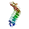 1h6tC 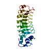 1d0bS S: Starting model for refinement C: citing same article ( |
|---|---|
| Similar structure data |


-Links
 PDBj
PDBj


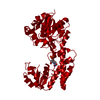
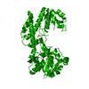
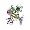
- Assembly
| Deposited unit | 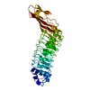
| ||||||||
|---|---|---|---|---|---|---|---|---|---|
| 1 | 
| ||||||||
| Unit cell |
|
-Components
| #1: Protein | Mass: 32585.611 Da / Num. of mol.: 1 / Fragment: LRR DOMAIN, RESIDUES 36-343 Source method: isolated from a genetically manipulated source Source: (gene. exp.) LISTERIA MONOCYTOGENES (bacteria) / Strain: EGD (SEROVAR 1/2A) / Plasmid: PGEX-6P-1 / Production host: ESCHERICHIA COLI (E. coli) / Strain (production host): BL21 / References: UniProt: Q9ZEY1 |
|---|---|
| #2: Water | ChemComp-HOH / Water Mass: 18.015 Da / Num. of mol.: 403 / Source method: isolated from a natural source / Formula: H2O Mass: 18.015 Da / Num. of mol.: 403 / Source method: isolated from a natural source / Formula: H2O |
| Sequence details | THE TWO N-TERMINAL RESIDUES (GLY SER) WERE INTRODUCED AS PART OF THE CLONING STRATEGY (PRESCISSION ...THE TWO N-TERMINAL RESIDUES (GLY SER) WERE INTRODUCED |
-Experimental details
-Experiment
| Experiment | Method: X-RAY DIFFRACTION / Number of used crystals: 1 |
|---|
- Sample preparation
| Crystal | Density Matthews: 2.17 Å3/Da / Density % sol: 42.8 % | ||||||||||||||||||||||||
|---|---|---|---|---|---|---|---|---|---|---|---|---|---|---|---|---|---|---|---|---|---|---|---|---|---|
| Crystal grow | Temperature: 293 K / pH: 5 Details: 10% PEG 8000, 0.1M TRIS PH 7.0, 0.2 M MGCL2. T=20C, PROTEIN CONC.=10 MG/ML | ||||||||||||||||||||||||
| Crystal grow | *PLUS pH: 7 / Method: sparse matrix method | ||||||||||||||||||||||||
| Components of the solutions | *PLUS
|
-Data collection
| Diffraction | Mean temperature: 100 K |
|---|---|
| Diffraction source | Source: ROTATING ANODE / Type: RIGAKU RU300 / Wavelength: 1.5418 |
| Detector | Type: RAXIS IV++ / Detector: IMAGE PLATE / Date: Jul 3, 2000 / Details: OSMIC CONFOCAL MIRRORS |
| Radiation | Protocol: SINGLE WAVELENGTH / Monochromatic (M) / Laue (L): M / Scattering type: x-ray |
| Radiation wavelength | Wavelength: 1.5418 Å / Relative weight: 1 |
| Reflection | Resolution: 1.6→61 Å / Num. obs: 37671 / % possible obs: 95.3 % / Observed criterion σ(I): 0 / Redundancy: 2 % / Rmerge(I) obs: 0.033 / Net I/σ(I): 22 |
| Reflection shell | Resolution: 1.6→1.66 Å / Redundancy: 1.9 % / Rmerge(I) obs: 0.162 / Mean I/σ(I) obs: 7.42 / % possible all: 91.2 |
| Reflection | *PLUS Highest resolution: 1.8 Å / Lowest resolution: 40 Å / Num. obs: 32149 / % possible obs: 98.4 % / Redundancy: 3.2 % / Rmerge(I) obs: 0.06 |
| Reflection shell | *PLUS Highest resolution: 1.8 Å / Lowest resolution: 1.86 Å / % possible obs: 96.5 % / Redundancy: 2.8 % / Rmerge(I) obs: 0.321 / Mean I/σ(I) obs: 3.3 |
- Processing
| Software |
| ||||||||||||||||||||||||||||||||||||||||||||||||||||||||||||||||||||||||||||||||||||||||||||||||||||||||||||||||||||||||||||||||||||||||||||||||||||||||||||||||||||||||||||||||||||||
|---|---|---|---|---|---|---|---|---|---|---|---|---|---|---|---|---|---|---|---|---|---|---|---|---|---|---|---|---|---|---|---|---|---|---|---|---|---|---|---|---|---|---|---|---|---|---|---|---|---|---|---|---|---|---|---|---|---|---|---|---|---|---|---|---|---|---|---|---|---|---|---|---|---|---|---|---|---|---|---|---|---|---|---|---|---|---|---|---|---|---|---|---|---|---|---|---|---|---|---|---|---|---|---|---|---|---|---|---|---|---|---|---|---|---|---|---|---|---|---|---|---|---|---|---|---|---|---|---|---|---|---|---|---|---|---|---|---|---|---|---|---|---|---|---|---|---|---|---|---|---|---|---|---|---|---|---|---|---|---|---|---|---|---|---|---|---|---|---|---|---|---|---|---|---|---|---|---|---|---|---|---|---|---|
| Refinement | Method to determine structure: MOLECULAR REPLACEMENT Starting model: PDB ENTRY 1D0B Resolution: 1.8→76.7 Å / Cor.coef. Fo:Fc: 0.963 / Cor.coef. Fo:Fc free: 0.941 / SU B: 3.536 / SU ML: 0.111 / Cross valid method: THROUGHOUT / ESU R: 0.115 / ESU R Free: 0.12 / Stereochemistry target values: MAXIMUM LIKELIHOOD / Details: HYDROGENS HAVE BEEN ADDED IN THE RIDING POSITIONS
| ||||||||||||||||||||||||||||||||||||||||||||||||||||||||||||||||||||||||||||||||||||||||||||||||||||||||||||||||||||||||||||||||||||||||||||||||||||||||||||||||||||||||||||||||||||||
| Solvent computation | Ion probe radii: 0.8 Å / Shrinkage radii: 0.8 Å / VDW probe radii: 1.4 Å / Solvent model: BABINET MODEL WITH MASK | ||||||||||||||||||||||||||||||||||||||||||||||||||||||||||||||||||||||||||||||||||||||||||||||||||||||||||||||||||||||||||||||||||||||||||||||||||||||||||||||||||||||||||||||||||||||
| Refinement step | Cycle: LAST / Resolution: 1.8→76.7 Å
| ||||||||||||||||||||||||||||||||||||||||||||||||||||||||||||||||||||||||||||||||||||||||||||||||||||||||||||||||||||||||||||||||||||||||||||||||||||||||||||||||||||||||||||||||||||||
| Refine LS restraints |
|