 Movie
Movie Controller


+ Open data

- Basic information
| Entry | Database: PDB / ID: 1h3i | ||||||
|---|---|---|---|---|---|---|---|
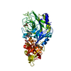
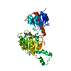
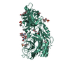
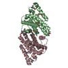
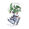
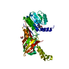
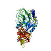
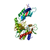
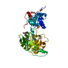
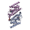
| Title | Crystal structure of the Histone Methyltransferase SET7/9 | ||||||
 Components Components | HISTONE H3 LYSINE 4 SPECIFIC METHYLTRANSFERASE | ||||||
 Keywords Keywords |  TRANSFERASE / METHYLTRANSFERASE TRANSFERASE / METHYLTRANSFERASE | ||||||
| Function / homology |  Function and homology information Function and homology informationheterochromatin organization / peptidyl-lysine monomethylation / peptidyl-lysine dimethylation / [histone H3]-lysine4 N-methyltransferase / histone H3K4 monomethyltransferase activity / protein-lysine N-methyltransferase activity / histone H3 methyltransferase activity / histone methyltransferase activity / PKMTs methylate histone lysines / p53 binding ...heterochromatin organization / peptidyl-lysine monomethylation / peptidyl-lysine dimethylation / [histone H3]-lysine4 N-methyltransferase / histone H3K4 monomethyltransferase activity / protein-lysine N-methyltransferase activity / histone H3 methyltransferase activity / histone methyltransferase activity / PKMTs methylate histone lysines / p53 binding / chromosome / chromatin organization / response to ethanol / DNA damage response / chromatin binding / nucleolus / positive regulation of DNA-templated transcription / nucleoplasm / nucleusSimilarity search - Function | ||||||
| Biological species |  HOMO SAPIENS (human) HOMO SAPIENS (human) | ||||||
| Method | X-RAY DIFFRACTION / SYNCHROTRON / MAD / Resolution: 2.1 Å | ||||||
 Authors Authors | Wilson, J.R. / Jing, C. / Walker, P.A. / Martin, S.R. / Howell, S.A. / Blackburn, G.M. / Gamblin, S.J. / Xiao, B. | ||||||
 Citation Citation | Journal: Cell(Cambridge,Mass.) / Year: 2002 Title: Crystal Structure and Functional Analysis of the Histone Methyltransferase Set7/9 Authors: Wilson, J.R. / Jing, C. / Walker, P.A. / Martin, S.R. / Howell, S.A. / Blackburn, G.M. / Gamblin, S.J. / Xiao, B. | ||||||
| History |
| ||||||
| Remark 700 | SHEET THE SHEET STRUCTURE OF THIS MOLECULE IS BIFURCATED. IN ORDER TO REPRESENT THIS FEATURE IN ... SHEET THE SHEET STRUCTURE OF THIS MOLECULE IS BIFURCATED. IN ORDER TO REPRESENT THIS FEATURE IN THE SHEET RECORDS BELOW, TWO SHEETS ARE DEFINED. |
- Structure visualization
| Structure viewer | Molecule: MolmilJmol/JSmol |
|---|
- Downloads & links
-Download
| PDBx/mmCIF format | 1h3i.cif.gz | 129.8 KB | Display | PDBx/mmCIF format |
|---|---|---|---|---|
| PDB format | pdb1h3i.ent.gz | 107 KB | Display | PDB format |
| PDBx/mmJSON format | 1h3i.json.gz | Tree view | PDBx/mmJSON format | |
| Others |  Other downloads Other downloads |
-Validation report
| Arichive directory | https://data.pdbj.org/pub/pdb/validation_reports/h3/1h3iftp://data.pdbj.org/pub/pdb/validation_reports/h3/1h3i | HTTPS FTP |
|---|
-Related structure data
| Similar structure data |
|---|
-Links
 PDBj
PDBj- Assembly
| Deposited unit | 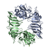
| ||||||||
|---|---|---|---|---|---|---|---|---|---|
| 1 | 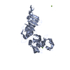
| ||||||||
| 2 | 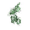
| ||||||||
| Unit cell |
|
-Components
| #1: Protein | Mass: 32571.914 Da / Num. of mol.: 2 / Fragment: N-DOMAIN, SET-DOMAIN, RESIDUES 52-344 Source method: isolated from a genetically manipulated source Source: (gene. exp.) HOMO SAPIENS (human) / Production host:  ESCHERICHIA COLI (E. coli) ESCHERICHIA COLI (E. coli)References: UniProt: Q8WTS6, histone-lysine N-methyltransferase#2: Chemical |   Mass: 24.305 Da / Num. of mol.: 3 / Source method: obtained synthetically / Formula: Mg Mass: 24.305 Da / Num. of mol.: 3 / Source method: obtained synthetically / Formula: Mg#3: Water | ChemComp-HOH / | Water Mass: 18.015 Da / Num. of mol.: 344 / Source method: isolated from a natural source / Formula: H2O Mass: 18.015 Da / Num. of mol.: 344 / Source method: isolated from a natural source / Formula: H2O |
|---|
-Experimental details
-Experiment
| Experiment | Method: X-RAY DIFFRACTION |
|---|
- Sample preparation
| Crystal | Density Matthews: 2.44 Å3/Da / Density % sol: 49.59 % | |||||||||||||||||||||||||||||||||||||||||||||||||
|---|---|---|---|---|---|---|---|---|---|---|---|---|---|---|---|---|---|---|---|---|---|---|---|---|---|---|---|---|---|---|---|---|---|---|---|---|---|---|---|---|---|---|---|---|---|---|---|---|---|---|
| Crystal grow | pH: 7 / Details: pH 7.00 | |||||||||||||||||||||||||||||||||||||||||||||||||
| Crystal grow | *PLUS Method: vapor diffusion | |||||||||||||||||||||||||||||||||||||||||||||||||
| Components of the solutions | *PLUS
|
-Data collection
| Diffraction | Mean temperature: 100 K | ||||||||||||
|---|---|---|---|---|---|---|---|---|---|---|---|---|---|
| Diffraction source | Source: SYNCHROTRON / Site: ESRF  / Beamline: ID14-4 / Wavelength: 0.9794, 0.9394, 0.9800 / Beamline: ID14-4 / Wavelength: 0.9794, 0.9394, 0.9800 | ||||||||||||
| Detector | Type: ADSC CCD / Detector: CCD | ||||||||||||
| Radiation | Protocol: MAD / Monochromatic (M) / Laue (L): M / Scattering type: x-ray | ||||||||||||
| Radiation wavelength |
| ||||||||||||
| Reflection | Resolution: 2.1→20 Å / Num. obs: 37053 / % possible obs: 95.4 % / Redundancy: 3.4 % / Rmerge(I) obs: 0.067 | ||||||||||||
| Reflection | *PLUS Lowest resolution: 30 Å / % possible obs: 84 % / Redundancy: 3.1 % / Rmerge(I) obs: 0.048 |
- Processing
| Software |
| ||||||||||||||||||||||||||||||||||||||||||||||||||||||||||||||||||||||||||||||||||||||||||||||||||||||||||||||||||||||||||||||||||||||||||||||||||||||||||||||||||||||||||||||||||||||
|---|---|---|---|---|---|---|---|---|---|---|---|---|---|---|---|---|---|---|---|---|---|---|---|---|---|---|---|---|---|---|---|---|---|---|---|---|---|---|---|---|---|---|---|---|---|---|---|---|---|---|---|---|---|---|---|---|---|---|---|---|---|---|---|---|---|---|---|---|---|---|---|---|---|---|---|---|---|---|---|---|---|---|---|---|---|---|---|---|---|---|---|---|---|---|---|---|---|---|---|---|---|---|---|---|---|---|---|---|---|---|---|---|---|---|---|---|---|---|---|---|---|---|---|---|---|---|---|---|---|---|---|---|---|---|---|---|---|---|---|---|---|---|---|---|---|---|---|---|---|---|---|---|---|---|---|---|---|---|---|---|---|---|---|---|---|---|---|---|---|---|---|---|---|---|---|---|---|---|---|---|---|---|---|
| Refinement | Method to determine structure: MAD / Resolution: 2.1→20 Å / Cor.coef. Fo:Fc: 0.94 / Cor.coef. Fo:Fc free: 0.917 / SU B: 5.105 / SU ML: 0.141 / Cross valid method: THROUGHOUT / ESU R: 0.263 / ESU R Free: 0.209 / Stereochemistry target values: MAXIMUM LIKELIHOOD
| ||||||||||||||||||||||||||||||||||||||||||||||||||||||||||||||||||||||||||||||||||||||||||||||||||||||||||||||||||||||||||||||||||||||||||||||||||||||||||||||||||||||||||||||||||||||
| Solvent computation | Ion probe radii: 0.8 Å / Shrinkage radii: 0.8 Å / VDW probe radii: 1.4 Å / Solvent model: BABINET MODEL WITH MASK | ||||||||||||||||||||||||||||||||||||||||||||||||||||||||||||||||||||||||||||||||||||||||||||||||||||||||||||||||||||||||||||||||||||||||||||||||||||||||||||||||||||||||||||||||||||||
| Displacement parameters | Biso mean: 19.9 Å2
| ||||||||||||||||||||||||||||||||||||||||||||||||||||||||||||||||||||||||||||||||||||||||||||||||||||||||||||||||||||||||||||||||||||||||||||||||||||||||||||||||||||||||||||||||||||||
| Refinement step | Cycle: LAST / Resolution: 2.1→20 Å
| ||||||||||||||||||||||||||||||||||||||||||||||||||||||||||||||||||||||||||||||||||||||||||||||||||||||||||||||||||||||||||||||||||||||||||||||||||||||||||||||||||||||||||||||||||||||
| Refine LS restraints |
|