 Movie
Movie Controller

[English] 日本語
 Yorodumi
- PDB-1h0o: Cobalt substitution of mouse R2 ribonucleotide reductase to model... -
+ Open data

- Basic information
| Entry | Database: PDB / ID: 1h0o | ||||||
|---|---|---|---|---|---|---|---|
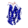
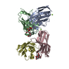
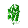
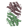
| Title | Cobalt substitution of mouse R2 ribonucleotide reductase to model the reactive diferrous state | ||||||
 Components Components | RIBONUCLEOSIDE-DIPHOSPHATE REDUCTASE Ribonucleotide reductase Ribonucleotide reductase | ||||||
 Keywords Keywords | OXIDOREDUCTASE / RIBONUCLEOTIDE REDUCTASE / DINUCLEAR METAL-CLUSTER | ||||||
| Function / homology |  Function and homology information Function and homology informationdeoxyribonucleotide metabolic process / Interconversion of nucleotide di- and triphosphates / ribonucleoside diphosphate metabolic process / 2'-deoxyribonucleotide biosynthetic process / ribonucleoside-diphosphate reductase complex / ribonucleoside-diphosphate reductase / ribonucleoside-diphosphate reductase activity, thioredoxin disulfide as acceptor / deoxyribonucleotide biosynthetic process / protein heterotetramerization / blastocyst development ...deoxyribonucleotide metabolic process / Interconversion of nucleotide di- and triphosphates / ribonucleoside diphosphate metabolic process / 2'-deoxyribonucleotide biosynthetic process / ribonucleoside-diphosphate reductase complex / ribonucleoside-diphosphate reductase / ribonucleoside-diphosphate reductase activity, thioredoxin disulfide as acceptor / deoxyribonucleotide biosynthetic process / protein heterotetramerization / blastocyst development / positive regulation of G1/S transition of mitotic cell cycle / ferric iron binding / nuclear envelope / protein homodimerization activity / identical protein binding / cytosolSimilarity search - Function | ||||||
| Biological species |  MUS MUSCULUS (house mouse) MUS MUSCULUS (house mouse) | ||||||
| Method | X-RAY DIFFRACTION / SYNCHROTRON / MOLECULAR REPLACEMENT / Resolution: 2.2 Å | ||||||
 Authors Authors | Strand, K.R. / Karlsen, S. / Andersson, K.K. | ||||||
 Citation Citation | Journal: J.Biol.Chem. / Year: 2002 Title: Cobalt Substitution of Mouse R2 Ribonucleotide Reductase as a Model for Thereactive Diferrous State. Spectroscopic and Structural Evidence for a Ferromagnetically Coupled Dinuclear Cobalt Cluster Authors: Strand, K.R. / Karlsen, S. / Andersson, K.K. | ||||||
| History |
|
- Structure visualization
| Structure viewer | Molecule: MolmilJmol/JSmol |
|---|
- Downloads & links
-Download
| PDBx/mmCIF format | 1h0o.cif.gz | 75.2 KB | Display | PDBx/mmCIF format |
|---|---|---|---|---|
| PDB format | pdb1h0o.ent.gz | 55.3 KB | Display | PDB format |
| PDBx/mmJSON format | 1h0o.json.gz | Tree view | PDBx/mmJSON format | |
| Others |  Other downloads Other downloads |
-Validation report
| Arichive directory | https://data.pdbj.org/pub/pdb/validation_reports/h0/1h0oftp://data.pdbj.org/pub/pdb/validation_reports/h0/1h0o | HTTPS FTP |
|---|
-Related structure data
| Related structure data |  1h0nC  1xsmS C: citing same article ( S: Starting model for refinement |
|---|---|
| Similar structure data |


-Links
 PDBj
PDBj

- Assembly
| Deposited unit | 
| ||||||||
|---|---|---|---|---|---|---|---|---|---|
| 1 | 
| ||||||||
| Unit cell |
|
-Components
| #1: Protein | Ribonucleotide reductase / RIBONUCLEOTIDE REDUCTASE SMALL CHAIN Mass: 45149.547 Da / Num. of mol.: 1 Source method: isolated from a genetically manipulated source Details: COBALT-SUBSTITUTED / Source: (gene. exp.) MUS MUSCULUS (house mouse) / Production host:  ESCHERICHIA COLI (E. coli) ESCHERICHIA COLI (E. coli)References: UniProt: P11157, ribonucleoside-diphosphate reductase |
|---|---|
| #2: Chemical | ChemComp-CO /   Mass: 58.933 Da / Num. of mol.: 1 / Source method: obtained synthetically / Formula: Co Mass: 58.933 Da / Num. of mol.: 1 / Source method: obtained synthetically / Formula: Co |
| #3: Water | ChemComp-HOH / Water Mass: 18.015 Da / Num. of mol.: 60 / Source method: isolated from a natural source / Formula: H2O Mass: 18.015 Da / Num. of mol.: 60 / Source method: isolated from a natural source / Formula: H2O |
| Compound details | DNA SYNTHESIS PRECURSOR PROVIDER |
-Experimental details
-Experiment
| Experiment | Method: X-RAY DIFFRACTION / Number of used crystals: 1 |
|---|
- Sample preparation
| Crystal | Density Matthews: 2.05 Å3/Da / Density % sol: 40.05 % | ||||||||||||||||||||||||||||
|---|---|---|---|---|---|---|---|---|---|---|---|---|---|---|---|---|---|---|---|---|---|---|---|---|---|---|---|---|---|
| Crystal grow | pH: 4.7 Details: 0.1 M NA-ACETATE BUFFER PH4.7, 1.2 M NACL, 7.5 MG/ML APO-R2 PROTEIN. CRYSTALS WERE MADE BY CO-CRYSTALLISATION (3.8 EQV. CO2+)., pH 4.70 | ||||||||||||||||||||||||||||
| Crystal grow | *PLUS Method: vapor diffusion, hanging drop | ||||||||||||||||||||||||||||
| Components of the solutions | *PLUS
|
-Data collection
| Diffraction | Mean temperature: 100 K |
|---|---|
| Diffraction source | Source: SYNCHROTRON / Site: ESRF  / Beamline: ID14-1 / Wavelength: 0.934 / Beamline: ID14-1 / Wavelength: 0.934 |
| Detector | Type: MARRESEARCH / Detector: CCD / Date: Mar 15, 2001 |
| Radiation | Protocol: SINGLE WAVELENGTH / Monochromatic (M) / Laue (L): M / Scattering type: x-ray |
| Radiation wavelength | Wavelength: 0.934 Å / Relative weight: 1 |
| Reflection | Resolution: 2.2→30 Å / Num. obs: 22045 / Biso Wilson estimate: 25.1 Å2 |
- Processing
| Software |
| ||||||||||||||||||||||||||||||||||||||||||||||||||||||||||||||||||||||||||||||||
|---|---|---|---|---|---|---|---|---|---|---|---|---|---|---|---|---|---|---|---|---|---|---|---|---|---|---|---|---|---|---|---|---|---|---|---|---|---|---|---|---|---|---|---|---|---|---|---|---|---|---|---|---|---|---|---|---|---|---|---|---|---|---|---|---|---|---|---|---|---|---|---|---|---|---|---|---|---|---|---|---|---|
| Refinement | Method to determine structure: MOLECULAR REPLACEMENT Starting model: PDB ENTRY 1XSM Resolution: 2.2→8 Å / Data cutoff high absF: 572486.45 / Isotropic thermal model: RESTRAINED / Cross valid method: THROUGHOUT
| ||||||||||||||||||||||||||||||||||||||||||||||||||||||||||||||||||||||||||||||||
| Solvent computation | Solvent model: FLAT MODEL / Bsol: 55.3783 Å2 / ksol: 0.384623 e/Å3 | ||||||||||||||||||||||||||||||||||||||||||||||||||||||||||||||||||||||||||||||||
| Displacement parameters | Biso mean: 45.6 Å2
| ||||||||||||||||||||||||||||||||||||||||||||||||||||||||||||||||||||||||||||||||
| Refine analyze |
| ||||||||||||||||||||||||||||||||||||||||||||||||||||||||||||||||||||||||||||||||
| Refinement step | Cycle: LAST / Resolution: 2.2→8 Å
| ||||||||||||||||||||||||||||||||||||||||||||||||||||||||||||||||||||||||||||||||
| Refine LS restraints |
| ||||||||||||||||||||||||||||||||||||||||||||||||||||||||||||||||||||||||||||||||
| LS refinement shell | Resolution: 2.2→2.34 Å / Rfactor Rfree error: 0.019 / Total num. of bins used: 6
| ||||||||||||||||||||||||||||||||||||||||||||||||||||||||||||||||||||||||||||||||
| Xplor file |
| ||||||||||||||||||||||||||||||||||||||||||||||||||||||||||||||||||||||||||||||||
| Refine LS restraints | *PLUS
|
