 Movie
Movie Controller

[English] 日本語
 Yorodumi
- PDB-1gyo: Crystal structure of the di-tetraheme cytochrome c3 from Desulfov... -
+ Open data

- Basic information
| Entry | Database: PDB / ID: 1gyo | ||||||
|---|---|---|---|---|---|---|---|
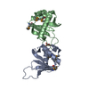
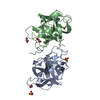
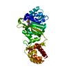
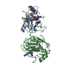
| Title | Crystal structure of the di-tetraheme cytochrome c3 from Desulfovibrio gigas at 1.2 Angstrom resolution | ||||||
 Components Components | CYTOCHROME C3, A DIMERIC CLASS III C-TYPE CYTOCHROME | ||||||
 Keywords Keywords |  ELECTRON TRANSPORT / CYTOCHROME C3 / DI-TETRAHEME / AB INITIO / ELECTRON TRANSFER ELECTRON TRANSPORT / CYTOCHROME C3 / DI-TETRAHEME / AB INITIO / ELECTRON TRANSFER | ||||||
| Function / homology |  Function and homology information Function and homology information | ||||||
| Biological species |  DESULFOVIBRIO GIGAS (bacteria) DESULFOVIBRIO GIGAS (bacteria) | ||||||
| Method | X-RAY DIFFRACTION / SYNCHROTRON / DIRECT METHODS / Resolution: 1.2 Å | ||||||
 Authors Authors | Aragao, D. / Frazao, C. / Sieker, L. / Sheldrick, G.M. / Legall, J. / Carrondo, M.A. | ||||||
 Citation Citation | Journal: Acta Crystallogr.,Sect.D / Year: 2003 Title: Structure of Dimeric Cytochrome C3 from Desulfovibrio Gigas at 1.2 A Resolution Authors: Aragao, D. / Frazao, C. / Sieker, L. / Sheldrick, G.M. / Legall, J. / Carrondo, M.A. #1: Journal: J.Biol.Inorg.Chem. / Year: 1999 Title: Ab Initio Structure Solution of a Dimeric Cytochrome C3 from Desulfovibrio Gigas Containing Disulfide Bridges Authors: Frazao, C. / Sieker, L. / Sheldrick, G.M. / Lamzin, V. / Legall, J. / Carrondo, M.A. #2: Journal: FEBS Lett. / Year: 1986 Title: Preliminary X-Ray Studies of the Tetra-Heme Cytochrome C3 and the Octa-Heme Cytochrome C3 from Desulfovibrio Gigas Authors: Sieker, L.C. / Jensen, L.H. / Legall, J. / Carrondo, M.A. | ||||||
| History |
|
- Structure visualization
| Structure viewer | Molecule: MolmilJmol/JSmol |
|---|
- Downloads & links
-Download
| PDBx/mmCIF format | 1gyo.cif.gz | 126.7 KB | Display | PDBx/mmCIF format |
|---|---|---|---|---|
| PDB format | pdb1gyo.ent.gz | 103.6 KB | Display | PDB format |
| PDBx/mmJSON format | 1gyo.json.gz | Tree view | PDBx/mmJSON format | |
| Others |  Other downloads Other downloads |
-Validation report
| Arichive directory | https://data.pdbj.org/pub/pdb/validation_reports/gy/1gyoftp://data.pdbj.org/pub/pdb/validation_reports/gy/1gyo | HTTPS FTP |
|---|
-Related structure data
| Similar structure data |
|---|


-Links
 PDBj
PDBj


- Assembly
| Deposited unit | 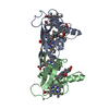
| ||||||||
|---|---|---|---|---|---|---|---|---|---|
| 1 |
| ||||||||
| Unit cell |
| ||||||||
| Noncrystallographic symmetry (NCS) | NCS oper: (Code: given Matrix: (-0.5378, 0.8381, 0.0914), Vector : |
-Components
| #1: Protein | Mass: 12240.867 Da / Num. of mol.: 2 / Source method: isolated from a natural source / Source: (natural) DESULFOVIBRIO GIGAS (bacteria) / References: UniProt: Q9R638#2: Chemical | ChemComp-HEC / Heme C  Mass: 618.503 Da / Num. of mol.: 8 / Source method: obtained synthetically / Formula: C34H34FeN4O4 Mass: 618.503 Da / Num. of mol.: 8 / Source method: obtained synthetically / Formula: C34H34FeN4O4#3: Chemical | Glycerol  Mass: 92.094 Da / Num. of mol.: 2 / Source method: obtained synthetically / Formula: C3H8O3 Mass: 92.094 Da / Num. of mol.: 2 / Source method: obtained synthetically / Formula: C3H8O3#4: Water | ChemComp-HOH / | Water Mass: 18.015 Da / Num. of mol.: 280 / Source method: isolated from a natural source / Formula: H2O Mass: 18.015 Da / Num. of mol.: 280 / Source method: isolated from a natural source / Formula: H2O |
|---|
-Experimental details
-Experiment
| Experiment | Method: X-RAY DIFFRACTION / Number of used crystals: 2 |
|---|
- Sample preparation
| Crystal | Density Matthews: 2.3 Å3/Da / Density % sol: 47.4 % |
|---|---|
| Crystal grow | pH: 6.5 Details: SALTING OUT FROM A PROTEIN SOLUTION IN TRIS/MALEATE PH=6.5, pH 6.50 |
-Data collection
| Diffraction | Mean temperature: 110 K |
|---|---|
| Diffraction source | Source: SYNCHROTRON / Site: EMBL/DESY, HAMBURG  / Beamline: BW7B / Wavelength: 1.1037 / Beamline: BW7B / Wavelength: 1.1037 |
| Detector | Type: MARRESEARCH / Detector: IMAGE PLATE / Date: May 15, 1997 / Details: MIRRORS |
| Radiation | Protocol: SINGLE WAVELENGTH / Monochromatic (M) / Laue (L): M / Scattering type: x-ray |
| Radiation wavelength | Wavelength: 1.1037 Å / Relative weight: 1 |
| Reflection | Resolution: 1.2→26.44 Å / Num. obs: 104411 / % possible obs: 98.7 % / Redundancy: 4.9 % / Rmerge(I) obs: 0.097 / Net I/σ(I): 16.3 |
| Reflection shell | Resolution: 1.2→1.21 Å / Redundancy: 2.5 % / Rmerge(I) obs: 0.437 / Mean I/σ(I) obs: 2.8 / % possible all: 95.6 |
| Reflection shell | *PLUS % possible obs: 95.6 % |
- Processing
| Software |
| |||||||||||||||||||||||||||||||||
|---|---|---|---|---|---|---|---|---|---|---|---|---|---|---|---|---|---|---|---|---|---|---|---|---|---|---|---|---|---|---|---|---|---|---|
| Refinement | Method to determine structure: DIRECT METHODS / Resolution: 1.2→26.5 Å / Num. parameters: 21173 / Num. restraintsaints: 50776 / Cross valid method: THROUGHOUT / σ(F): 0 StereochEM target val spec case: HEME GROUPS WERE RESTRAINED TO BE SIMILAR BUT WITHOUT TARGET VALUES, DUE TO THEIR INTERNAL SYMMETRY AND FOUR- OR EIGHT-FOLD REDUNDANCY Stereochemistry target values: ENGH & HUBER Details: C-TERMINI RESIDUES ALA107-GLN108- LYS109 ARE MISSING IN EACH MONOMER AS THEY ARE NOT VISIBLE IN ELECTRON DENSITY MAPS. SOLVENT WATER MOLECULES THAT FOLLOW THE INTER-MONOMERS NCS RELATIONSHIP ...Details: C-TERMINI RESIDUES ALA107-GLN108- LYS109 ARE MISSING IN EACH MONOMER AS THEY ARE NOT VISIBLE IN ELECTRON DENSITY MAPS. SOLVENT WATER MOLECULES THAT FOLLOW THE INTER-MONOMERS NCS RELATIONSHIP ARE DIVIDED INTO TWO CHAINS, X FOR THOSE CLOSE TO MONOMER A AND Y FOR THOSE CLOSE TO MONOMER B. OTHER SOLVENT MOLECULES ARE ASSIGNED TO CHAIN Z. THE GLYCEROL MOLECULES IN THE STRUCTURE ARE ARTIFACTS OF THE CRYO- PROTECTANT.
| |||||||||||||||||||||||||||||||||
| Solvent computation | Solvent model: MOEWS & KRETSINGER | |||||||||||||||||||||||||||||||||
| Refine analyze | Num. disordered residues: 19 / Occupancy sum non hydrogen: 2286 | |||||||||||||||||||||||||||||||||
| Refinement step | Cycle: LAST / Resolution: 1.2→26.5 Å
| |||||||||||||||||||||||||||||||||
| Refine LS restraints |
| |||||||||||||||||||||||||||||||||
| Software | *PLUS Name: SHELXL / Version: 97 / Classification: refinement | |||||||||||||||||||||||||||||||||
| Refinement | *PLUS Highest resolution: 1.2 Å / Rfactor obs: 0.124 / Rfactor Rfree: 0.148 / Rfactor Rwork: 0.124 | |||||||||||||||||||||||||||||||||
| Solvent computation | *PLUS | |||||||||||||||||||||||||||||||||
| Displacement parameters | *PLUS |
