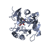
 Movie
Movie Controller


+ Open data

- Basic information
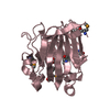
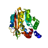
| Entry | Database: PDB / ID: 1gv9 | ||||||
|---|---|---|---|---|---|---|---|
| Title | p58/ERGIC-53 | ||||||
 Components Components | P58/ERGIC-53 | ||||||
 Keywords Keywords |  CARBOHYDRATE BINDING / LECTIN CARBOHYDRATE BINDING / LECTIN | ||||||
| Function / homology |  Function and homology information Function and homology informationRHOD GTPase cycle / : / RHOG GTPase cycle / RAC2 GTPase cycle / : / RHOA GTPase cycle / Cargo concentration in the ER / positive regulation of organelle organization / COPII-mediated vesicle transport / endoplasmic reticulum organization ...RHOD GTPase cycle / : / RHOG GTPase cycle / RAC2 GTPase cycle / : / RHOA GTPase cycle / Cargo concentration in the ER / positive regulation of organelle organization / COPII-mediated vesicle transport / endoplasmic reticulum organization / COPII-coated ER to Golgi transport vesicle / Golgi organization / mannose binding / endoplasmic reticulum-Golgi intermediate compartment / endoplasmic reticulum to Golgi vesicle-mediated transport / endoplasmic reticulum-Golgi intermediate compartment membrane / sarcomere / protein transport / Golgi membrane / calcium ion binding / endoplasmic reticulum membrane / Golgi apparatus / endoplasmic reticulum / identical protein binding / metal ion bindingSimilarity search - Function | ||||||
| Biological species |  RATTUS NORVEGICUS (Norway rat) RATTUS NORVEGICUS (Norway rat) | ||||||
| Method | X-RAY DIFFRACTION / SYNCHROTRON / MIR / Resolution: 1.46 Å | ||||||
 Authors Authors | Velloso, L.M. / Svensson, K. / Schneider, G. / Pettersson, R.F. / Lindqvist, Y. | ||||||
 Citation Citation | Journal: J.Biol.Chem. / Year: 2002 Title: Crystal Structure of the Carbohydrate Recognition Domain of P58/Ergic-53, a Protein Involved in Glycoprotein Export from the Endoplasmic Reticulum. Authors: Velloso, L.M. / Svensson, K. / Schneider, G. / Pettersson, R.F. / Lindqvist, Y. | ||||||
| History |
| ||||||
| Remark 700 | SHEET THE SHEET STRUCTURE OF THIS MOLECULE IS BIFURCATED. IN ORDER TO REPRESENT THIS FEATURE IN ... SHEET THE SHEET STRUCTURE OF THIS MOLECULE IS BIFURCATED. IN ORDER TO REPRESENT THIS FEATURE IN THE SHEET RECORDS BELOW, TWO SHEETS ARE DEFINED. |
- Structure visualization
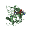
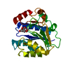
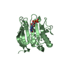
| Structure viewer | Molecule: MolmilJmol/JSmol |
|---|
- Downloads & links
-Download
| PDBx/mmCIF format | 1gv9.cif.gz | 57.8 KB | Display | PDBx/mmCIF format |
|---|---|---|---|---|
| PDB format | pdb1gv9.ent.gz | 44.5 KB | Display | PDB format |
| PDBx/mmJSON format | 1gv9.json.gz | Tree view | PDBx/mmJSON format | |
| Others |  Other downloads Other downloads |
-Validation report
| Arichive directory | https://data.pdbj.org/pub/pdb/validation_reports/gv/1gv9ftp://data.pdbj.org/pub/pdb/validation_reports/gv/1gv9 | HTTPS FTP |
|---|
-Related structure data
| Similar structure data |
|---|


-Links
 PDBj
PDBj- Assembly
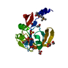
| Deposited unit | 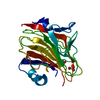
| ||||||||
|---|---|---|---|---|---|---|---|---|---|
| 1 |
| ||||||||
| Unit cell |
|
-Components
| #1: Protein | Mass: 28301.328 Da / Num. of mol.: 1 / Fragment: CARBOHYDRATE RECOGNITION DOMAIN / Source method: isolated from a natural source / Source: (natural) RATTUS NORVEGICUS (Norway rat) / References: UniProt: Q62902 | ||
|---|---|---|---|
| #2: Chemical | Sulfate  Mass: 96.063 Da / Num. of mol.: 2 / Source method: obtained synthetically / Formula: SO4 Mass: 96.063 Da / Num. of mol.: 2 / Source method: obtained synthetically / Formula: SO4#3: Water | ChemComp-HOH / | Water Mass: 18.015 Da / Num. of mol.: 191 / Source method: isolated from a natural source / Formula: H2O Mass: 18.015 Da / Num. of mol.: 191 / Source method: isolated from a natural source / Formula: H2O |
-Experimental details
-Experiment
| Experiment | Method: X-RAY DIFFRACTION |
|---|
- Sample preparation
| Crystal | Density Matthews: 2.4 Å3/Da / Density % sol: 48 % | |||||||||||||||||||||||||||||||||||||||||||||||||
|---|---|---|---|---|---|---|---|---|---|---|---|---|---|---|---|---|---|---|---|---|---|---|---|---|---|---|---|---|---|---|---|---|---|---|---|---|---|---|---|---|---|---|---|---|---|---|---|---|---|---|
| Crystal grow | pH: 7.25 / Details: pH 7.25 | |||||||||||||||||||||||||||||||||||||||||||||||||
| Crystal grow | *PLUS Method: vapor diffusion, hanging drop | |||||||||||||||||||||||||||||||||||||||||||||||||
| Components of the solutions | *PLUS
|
-Data collection
| Diffraction | Mean temperature: 110 K |
|---|---|
| Diffraction source | Source: SYNCHROTRON / Site: MAX II  / Beamline: I711 / Wavelength: 1.5418 / Beamline: I711 / Wavelength: 1.5418 |
| Detector | Type: MARRESEARCH |
| Radiation | Protocol: SINGLE WAVELENGTH / Monochromatic (M) / Laue (L): M / Scattering type: x-ray |
| Radiation wavelength | Wavelength: 1.5418 Å / Relative weight: 1 |
| Reflection | Resolution: 1.46→25 Å / Num. obs: 44706 / % possible obs: 95.6 % / Redundancy: 4.5 % / Rmerge(I) obs: 0.041 / Net I/σ(I): 22.2 |
| Reflection shell | *PLUS Highest resolution: 1.46 Å / Lowest resolution: 1.51 Å / % possible obs: 84.6 % / Rmerge(I) obs: 0.119 / Mean I/σ(I) obs: 7.3 |
- Processing
| Software |
| ||||||||||||||||||||||||||||||||||||||||||||||||||||||||||||||||||||||||||||||||||||||||||||||||||||||||||||||||||||||||||||||||||||||||||||||||||||||||||||||||||||||||||||||||||||||
|---|---|---|---|---|---|---|---|---|---|---|---|---|---|---|---|---|---|---|---|---|---|---|---|---|---|---|---|---|---|---|---|---|---|---|---|---|---|---|---|---|---|---|---|---|---|---|---|---|---|---|---|---|---|---|---|---|---|---|---|---|---|---|---|---|---|---|---|---|---|---|---|---|---|---|---|---|---|---|---|---|---|---|---|---|---|---|---|---|---|---|---|---|---|---|---|---|---|---|---|---|---|---|---|---|---|---|---|---|---|---|---|---|---|---|---|---|---|---|---|---|---|---|---|---|---|---|---|---|---|---|---|---|---|---|---|---|---|---|---|---|---|---|---|---|---|---|---|---|---|---|---|---|---|---|---|---|---|---|---|---|---|---|---|---|---|---|---|---|---|---|---|---|---|---|---|---|---|---|---|---|---|---|---|
| Refinement | Method to determine structure: MIR / Resolution: 1.46→23.64 Å / Cor.coef. Fo:Fc: 0.952 / Cor.coef. Fo:Fc free: 0.942 / SU B: 1.402 / SU ML: 0.056 / TLS residual ADP flag: LIKELY RESIDUAL / Cross valid method: THROUGHOUT / ESU R: 0.067 / ESU R Free: 0.068 / Stereochemistry target values: MAXIMUM LIKELIHOOD / Details: HYDROGENS HAVE BEEN ADDED IN THE RIDING POSITIONS
| ||||||||||||||||||||||||||||||||||||||||||||||||||||||||||||||||||||||||||||||||||||||||||||||||||||||||||||||||||||||||||||||||||||||||||||||||||||||||||||||||||||||||||||||||||||||
| Solvent computation | Ion probe radii: 0.8 Å / Shrinkage radii: 0.8 Å / VDW probe radii: 1.4 Å / Solvent model: BABINET MODEL WITH MASK | ||||||||||||||||||||||||||||||||||||||||||||||||||||||||||||||||||||||||||||||||||||||||||||||||||||||||||||||||||||||||||||||||||||||||||||||||||||||||||||||||||||||||||||||||||||||
| Refinement step | Cycle: LAST / Resolution: 1.46→23.64 Å
| ||||||||||||||||||||||||||||||||||||||||||||||||||||||||||||||||||||||||||||||||||||||||||||||||||||||||||||||||||||||||||||||||||||||||||||||||||||||||||||||||||||||||||||||||||||||
| Refine LS restraints |
|