 Movie
Movie Controller

[English] 日本語
 Yorodumi
- PDB-1gt9: High resolution crystal structure of a thermostable serine-carbox... -
+ Open data

- Basic information
| Entry | Database: PDB / ID: 1gt9 | ||||||
|---|---|---|---|---|---|---|---|
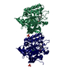
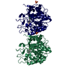
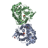
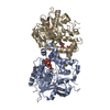
| Title | High resolution crystal structure of a thermostable serine-carboxyl type proteinase, kumamolisin (kscp) | ||||||
 Components Components | KUMAMOLYSIN | ||||||
 Keywords Keywords |  HYDROLASE / SERINE-CARBOXYL TYPE PROTEINASE / THERMOSTABLE HYDROLASE / SERINE-CARBOXYL TYPE PROTEINASE / THERMOSTABLE | ||||||
| Function / homology |  Function and homology information Function and homology information | ||||||
| Biological species |  BACILLUS SUBTILIS (bacteria) BACILLUS SUBTILIS (bacteria) | ||||||
| Method | X-RAY DIFFRACTION / SYNCHROTRON / MOLECULAR REPLACEMENT / Resolution: 1.38 Å | ||||||
 Authors Authors | Comellas-Bigler, M. / Fuentes-Prior, P. / Maskos, K. / Huber, R. / Oyama, H. / Uchida, K. / Dunn, B.M. / Oda, K. / Bode, W. | ||||||
 Citation Citation | Journal: Structure / Year: 2002 Title: The 1.4 A Crystal Structure of Kumamolysin. A Thermostable Serine-Carboxyl-Type Proteinase Authors: Comellas-Bigler, M. / Fuentes-Prior, P. / Maskos, K. / Huber, R. / Oyama, H. / Uchida, K. / Dunn, B.M. / Oda, K. / Bode, W. | ||||||
| History |
| ||||||
| Remark 700 | SHEET THE SHEET STRUCTURE OF THIS MOLECULE IS BIFURCATED. IN ORDER TO REPRESENT THIS FEATURE IN ... SHEET THE SHEET STRUCTURE OF THIS MOLECULE IS BIFURCATED. IN ORDER TO REPRESENT THIS FEATURE IN THE SHEET RECORDS BELOW, TWO SHEETS ARE DEFINED. |
- Structure visualization
| Structure viewer | Molecule: MolmilJmol/JSmol |
|---|
- Downloads & links
-Download
| PDBx/mmCIF format | 1gt9.cif.gz | 158.4 KB | Display | PDBx/mmCIF format |
|---|---|---|---|---|
| PDB format | pdb1gt9.ent.gz | 123.1 KB | Display | PDB format |
| PDBx/mmJSON format | 1gt9.json.gz | Tree view | PDBx/mmJSON format | |
| Others |  Other downloads Other downloads |
-Validation report
| Arichive directory | https://data.pdbj.org/pub/pdb/validation_reports/gt/1gt9ftp://data.pdbj.org/pub/pdb/validation_reports/gt/1gt9 | HTTPS FTP |
|---|
-Related structure data
| Related structure data | 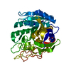 1gtgC 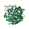 1gtjC 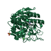 1gtlC  1ga1S C: citing same article ( S: Starting model for refinement |
|---|---|
| Similar structure data |


-Links
 PDBj
PDBj

- Assembly
| Deposited unit | 
| ||||||||
|---|---|---|---|---|---|---|---|---|---|
| 1 |
| ||||||||
| Unit cell |
|
-Components
| #1: Protein | Mass: 36317.012 Da / Num. of mol.: 2 Source method: isolated from a genetically manipulated source Source: (gene. exp.) BACILLUS SUBTILIS (bacteria)Description: BACTERIUM FOUND IN HOTSPRING WATER NEAR VOLCANOES Plasmid: PS3-A1 / Production host: ESCHERICHIA COLI (E. coli) / Strain (production host): JM 109 / References: UniProt: Q8RR56*PLUS#2: Chemical |   Mass: 40.078 Da / Num. of mol.: 2 / Source method: obtained synthetically / Formula: Ca Mass: 40.078 Da / Num. of mol.: 2 / Source method: obtained synthetically / Formula: Ca#3: Chemical | Sulfate  Mass: 96.063 Da / Num. of mol.: 2 / Source method: obtained synthetically / Formula: SO4 Mass: 96.063 Da / Num. of mol.: 2 / Source method: obtained synthetically / Formula: SO4#4: Water | ChemComp-HOH / | Water Mass: 18.015 Da / Num. of mol.: 566 / Source method: isolated from a natural source / Formula: H2O Mass: 18.015 Da / Num. of mol.: 566 / Source method: isolated from a natural source / Formula: H2O |
|---|
-Experimental details
-Experiment
| Experiment | Method: X-RAY DIFFRACTION / Number of used crystals: 1 |
|---|
- Sample preparation
| Crystal | Density Matthews: 2.17 Å3/Da / Density % sol: 43.39 % | ||||||||||||||||||||||||||||||||||||||||||
|---|---|---|---|---|---|---|---|---|---|---|---|---|---|---|---|---|---|---|---|---|---|---|---|---|---|---|---|---|---|---|---|---|---|---|---|---|---|---|---|---|---|---|---|
| Crystal grow | pH: 4.5 Details: 100 MM SODIUM ACETATE PH 4.5, 400 MM AMMONIUM SULPHATE | ||||||||||||||||||||||||||||||||||||||||||
| Crystal grow | *PLUS Temperature: 20 ℃ / pH: 5 / Method: vapor diffusion, sitting drop | ||||||||||||||||||||||||||||||||||||||||||
| Components of the solutions | *PLUS
|
-Data collection
| Diffraction | Mean temperature: 100 K |
|---|---|
| Diffraction source | Source: SYNCHROTRON / Site: MPG/DESY, HAMBURG  / Beamline: BW6 / Wavelength: 1.05 / Beamline: BW6 / Wavelength: 1.05 |
| Detector | Type: MARRESEARCH / Detector: CCD |
| Radiation | Protocol: SINGLE WAVELENGTH / Monochromatic (M) / Laue (L): M / Scattering type: x-ray |
| Radiation wavelength | Wavelength: 1.05 Å / Relative weight: 1 |
| Reflection | Resolution: 1.38→19.52 Å / Num. obs: 603967 / % possible obs: 95.5 % / Redundancy: 4.8 % / Rmerge(I) obs: 0.084 |
| Reflection shell | Resolution: 1.38→1.41 Å / Redundancy: 4.5 % / Rmerge(I) obs: 0.232 / % possible all: 79 |
| Reflection | *PLUS Num. obs: 120406 / Num. measured all: 603967 / Rmerge(I) obs: 0.064 |
| Reflection shell | *PLUS % possible obs: 79 % |
- Processing
| Software |
| ||||||||||||||||||||||||||||||||||||||||||||||||||||||||||||
|---|---|---|---|---|---|---|---|---|---|---|---|---|---|---|---|---|---|---|---|---|---|---|---|---|---|---|---|---|---|---|---|---|---|---|---|---|---|---|---|---|---|---|---|---|---|---|---|---|---|---|---|---|---|---|---|---|---|---|---|---|---|
| Refinement | Method to determine structure: MOLECULAR REPLACEMENT Starting model: PDB ENTRY 1GA1 Resolution: 1.38→19.52 Å / Data cutoff high absF: 10000 / Cross valid method: THROUGHOUT / σ(F): 0
| ||||||||||||||||||||||||||||||||||||||||||||||||||||||||||||
| Refinement step | Cycle: LAST / Resolution: 1.38→19.52 Å
| ||||||||||||||||||||||||||||||||||||||||||||||||||||||||||||
| Refine LS restraints |
| ||||||||||||||||||||||||||||||||||||||||||||||||||||||||||||
| Xplor file |
| ||||||||||||||||||||||||||||||||||||||||||||||||||||||||||||
| Refinement | *PLUS Num. reflection obs: 108310 | ||||||||||||||||||||||||||||||||||||||||||||||||||||||||||||
| Solvent computation | *PLUS | ||||||||||||||||||||||||||||||||||||||||||||||||||||||||||||
| Displacement parameters | *PLUS |
