 Movie
Movie Controller

[English] 日本語
 Yorodumi
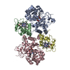
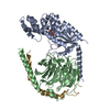
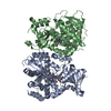
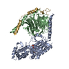
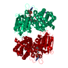
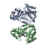
- PDB-1got: HETEROTRIMERIC COMPLEX OF A GT-ALPHA/GI-ALPHA CHIMERA AND THE GT-... -
+ Open data

- Basic information
| Entry | Database: PDB / ID: 1got | ||||||
|---|---|---|---|---|---|---|---|
| Title | HETEROTRIMERIC COMPLEX OF A GT-ALPHA/GI-ALPHA CHIMERA AND THE GT-BETA-GAMMA SUBUNITS | ||||||
 Components Components |
| ||||||
 Keywords Keywords | COMPLEX (GTP-BINDING/TRANSDUCER) / COMPLEX (GTP-BINDING-TRANSDUCER) /  G PROTEIN / HETEROTRIMER SIGNAL TRANSDUCTION / COMPLEX (GTP-BINDING-TRANSDUCER) complex G PROTEIN / HETEROTRIMER SIGNAL TRANSDUCTION / COMPLEX (GTP-BINDING-TRANSDUCER) complex | ||||||
| Function / homology |  Function and homology information Function and homology informationnegative regulation of cyclic-nucleotide phosphodiesterase activity / Olfactory Signaling Pathway / Sensory perception of sweet, bitter, and umami (glutamate) taste / Synthesis, secretion, and inactivation of Glucagon-like Peptide-1 (GLP-1) / eye photoreceptor cell development / G protein-coupled receptor complex / Inactivation, recovery and regulation of the phototransduction cascade / Activation of the phototransduction cascade / Activation of G protein gated Potassium channels / G-protein activation ...negative regulation of cyclic-nucleotide phosphodiesterase activity / Olfactory Signaling Pathway / Sensory perception of sweet, bitter, and umami (glutamate) taste / Synthesis, secretion, and inactivation of Glucagon-like Peptide-1 (GLP-1) / eye photoreceptor cell development / G protein-coupled receptor complex / Inactivation, recovery and regulation of the phototransduction cascade / Activation of the phototransduction cascade / Activation of G protein gated Potassium channels / G-protein activation / G beta:gamma signalling through PI3Kgamma / Prostacyclin signalling through prostacyclin receptor / Adrenaline,noradrenaline inhibits insulin secretion / G beta:gamma signalling through PLC beta / ADP signalling through P2Y purinoceptor 1 / Thromboxane signalling through TP receptor / Presynaptic function of Kainate receptors / G beta:gamma signalling through CDC42 / Inhibition of voltage gated Ca2+ channels via Gbeta/gamma subunits / Glucagon-type ligand receptors / acyl binding / G alpha (12/13) signalling events / G beta:gamma signalling through BTK / ADP signalling through P2Y purinoceptor 12 / Cooperation of PDCL (PhLP1) and TRiC/CCT in G-protein beta folding / Thrombin signalling through proteinase activated receptors (PARs) / Ca2+ pathway / Extra-nuclear estrogen signaling / G alpha (z) signalling events / G alpha (s) signalling events / G alpha (q) signalling events / photoreceptor outer segment membrane / G alpha (i) signalling events / Glucagon-like Peptide-1 (GLP1) regulates insulin secretion / Vasopressin regulates renal water homeostasis via Aquaporins / response to light stimulus / phototransduction / visual perception / photoreceptor inner segment / G protein-coupled receptor binding / G-protein beta/gamma-subunit complex binding / adenylate cyclase-modulating G protein-coupled receptor signaling pathway / protein localization / photoreceptor disc membrane / cellular response to catecholamine stimulus / adenylate cyclase-activating dopamine receptor signaling pathway / cellular response to prostaglandin E stimulus / sensory perception of taste / GDP binding / G-protein beta-subunit binding / heterotrimeric G-protein complex / signaling receptor complex adaptor activity / retina development in camera-type eye / GTPase binding / phospholipase C-activating G protein-coupled receptor signaling pathway / cell population proliferation / G protein-coupled receptor signaling pathway / GTPase activity / protein-containing complex binding / GTP binding / protein kinase binding / membrane / metal ion binding / cytoplasmSimilarity search - Function | ||||||
| Biological species |  Bos taurus (cattle) Bos taurus (cattle) | ||||||
| Method | X-RAY DIFFRACTION / SYNCHROTRON / MIR / Resolution: 2 Å | ||||||
 Authors Authors | Lambright, D.G. / Sondek, J. / Bohm, A. / Skiba, N.P. / Hamm, H.E. / Sigler, P.B. | ||||||
 Citation Citation | Journal: Nature / Year: 1996 Title: The 2.0 A crystal structure of a heterotrimeric G protein. Authors: Lambright, D.G. / Sondek, J. / Bohm, A. / Skiba, N.P. / Hamm, H.E. / Sigler, P.B. #1: Journal: Nature / Year: 1996Title: Crystal Structure of a Ga Protein Beta Gamma Dimer at 2.1A Resolution Authors: Sondek, J. / Bohm, A. / Lambright, D.G. / Hamm, H.E. / Sigler, P.B. #2: Journal: Nature / Year: 1994Title: Structural Determinants for Activation of the Alpha-Subunit of a Heterotrimeric G Protein Authors: Lambright, D.G. / Noel, J.P. / Hamm, H.E. / Sigler, P.B. #3: Journal: Nature / Year: 1994Title: Gtpase Mechanism of Gproteins from the 1.7-A Crystal Structure of Transducin Alpha-Gdp-Aif-4 Authors: Sondek, J. / Lambright, D.G. / Noel, J.P. / Hamm, H.E. / Sigler, P.B. #4: Journal: Nature / Year: 1993Title: The 2.2 A Crystal Structure of Transducin-Alpha Complexed with GTP Gamma S Authors: Noel, J.P. / Hamm, H.E. / Sigler, P.B. | ||||||
| History |
|
- Structure visualization
| Structure viewer | Molecule: MolmilJmol/JSmol |
|---|
- Downloads & links
-Download
| PDBx/mmCIF format | 1got.cif.gz | 169.5 KB | Display | PDBx/mmCIF format |
|---|---|---|---|---|
| PDB format | pdb1got.ent.gz | 137.2 KB | Display | PDB format |
| PDBx/mmJSON format | 1got.json.gz | Tree view | PDBx/mmJSON format | |
| Others |  Other downloads Other downloads |
-Validation report
| Arichive directory | https://data.pdbj.org/pub/pdb/validation_reports/go/1gotftp://data.pdbj.org/pub/pdb/validation_reports/go/1got | HTTPS FTP |
|---|
-Related structure data
| Similar structure data |
|---|


-Links
 PDBj
PDBj


- Assembly
| Deposited unit | 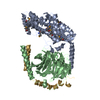
| |||||||||
|---|---|---|---|---|---|---|---|---|---|---|
| 1 |
| |||||||||
| Unit cell |
| |||||||||
| Components on special symmetry positions |
|
-Components
| #1: Protein | Mass: 40685.473 Da / Num. of mol.: 1 Source method: isolated from a genetically manipulated source Details: THIS IS A CHIMERIC PROTEIN WHERE RESIDUES 216-294 OF BOVINE GT ALPHA HAVE BEEN REPLACED WITH RESIDUES 220-298 OF RAT GI ALPHA 1 Source: (gene. exp.) Bos taurus (cattle) / Tissue: RETINA / Description: T7 LAC PROMOTER / Cell: ROD / Fragment: 216 - 294 / Organ: EYE / Organelle: ROD OUTER SEGMENTRod cell / Plasmid: PET VECTOR / Production host:  Escherichia coli (E. coli) / Strain (production host): PET / References: UniProt: P04695 Escherichia coli (E. coli) / Strain (production host): PET / References: UniProt: P04695 |
|---|---|
| #2: Protein | Mass: 37430.957 Da / Num. of mol.: 1 Source method: isolated from a genetically manipulated source Source: (gene. exp.) Bos taurus (cattle) / Tissue: RETINA / Cell: ROD / Organ: EYE / Organelle: ROD OUTER SEGMENTRod cell / References: UniProt: P62871 |
| #3: Protein | Mass: 8578.832 Da / Num. of mol.: 1 Source method: isolated from a genetically manipulated source Details: GT-BETA-GAMMA WAS TREATED WITH ENDOPROTEASE-LYSC TO REMOVE THE THREE C-TERMINAL AMINO ACIDS AND THE TERMINAL FARNESYL MOIETY OF GAMMA Source: (gene. exp.) Bos taurus (cattle) / Tissue: RETINA / Cell: ROD / Organ: EYE / Organelle: ROD OUTER SEGMENTRod cell / References: UniProt: P02698 |
| #4: Chemical | ChemComp-GDP / Guanosine diphosphate  Type: RNA linking / Mass: 443.201 Da / Num. of mol.: 1 / Source method: obtained synthetically / Formula: C10H15N5O11P2 / Comment: GDP, energy-carrying molecule*YM Type: RNA linking / Mass: 443.201 Da / Num. of mol.: 1 / Source method: obtained synthetically / Formula: C10H15N5O11P2 / Comment: GDP, energy-carrying molecule*YM |
| #5: Water | ChemComp-HOH / Water Mass: 18.015 Da / Num. of mol.: 616 / Source method: isolated from a natural source / Formula: H2O Mass: 18.015 Da / Num. of mol.: 616 / Source method: isolated from a natural source / Formula: H2O |
| Source details | GT-BETA-GAMMA WAS TREATED WITH ENDOPROTEASE-LYSC TO REMOVE THE THREE C-TERMINAL AMINO ACIDS AND THE ...GT-BETA-GAMMA WAS TREATED WITH ENDOPROTEA |
-Experimental details
-Experiment
| Experiment | Method: X-RAY DIFFRACTION / Number of used crystals: 1 |
|---|
- Sample preparation
| Crystal | Density Matthews: 2.53 Å3/Da / Density % sol: 52 % | ||||||||||||||||||||||||||||||||||||||||||||||||||||||||||||||||||||||||
|---|---|---|---|---|---|---|---|---|---|---|---|---|---|---|---|---|---|---|---|---|---|---|---|---|---|---|---|---|---|---|---|---|---|---|---|---|---|---|---|---|---|---|---|---|---|---|---|---|---|---|---|---|---|---|---|---|---|---|---|---|---|---|---|---|---|---|---|---|---|---|---|---|---|
| Crystal grow | Temperature: 277 K / Method: vapor diffusion, hanging drop / pH: 8 Details: 10 MG/ML OF HETEROTRIMERIC COMPLEX WERE MIXED 1:1 WITH WELL SOLUTION CONTAINING 10% PEG-8000, 50 MM TRIS, PH 8.0, 10% GLYCEROL, 50 MM NACL, .1 MM MERCAPTOETHANOL. MIXTURE EQUILIBRATED VS ...Details: 10 MG/ML OF HETEROTRIMERIC COMPLEX WERE MIXED 1:1 WITH WELL SOLUTION CONTAINING 10% PEG-8000, 50 MM TRIS, PH 8.0, 10% GLYCEROL, 50 MM NACL, .1 MM MERCAPTOETHANOL. MIXTURE EQUILIBRATED VS WELL SOLUTION IN HANGING DROPS AT 4 DEGREES C., vapor diffusion - hanging drop, temperature 277K | ||||||||||||||||||||||||||||||||||||||||||||||||||||||||||||||||||||||||
| Crystal grow | *PLUS Temperature: 4 ℃ / Method: vapor diffusion, hanging drop | ||||||||||||||||||||||||||||||||||||||||||||||||||||||||||||||||||||||||
| Components of the solutions | *PLUS
|
-Data collection
| Diffraction | Mean temperature: 100 K |
|---|---|
| Diffraction source | Source: SYNCHROTRON / Site: NSLS  / Beamline: X25 / Wavelength: 0.98 / Beamline: X25 / Wavelength: 0.98 |
| Detector | Type: MARRESEARCH / Detector: IMAGE PLATE / Date: May 10, 1995 |
| Radiation | Monochromatic (M) / Laue (L): M / Scattering type: x-ray |
| Radiation wavelength | Wavelength: 0.98 Å / Relative weight: 1 |
| Reflection | Resolution: 2→20 Å / Num. obs: 54174 / % possible obs: 99 % / Observed criterion σ(I): -3 / Redundancy: 4 % / Rmerge(I) obs: 0.061 |
- Processing
| Software |
| ||||||||||||||||||||||||||||||||||||||||||||||||||||||||||||
|---|---|---|---|---|---|---|---|---|---|---|---|---|---|---|---|---|---|---|---|---|---|---|---|---|---|---|---|---|---|---|---|---|---|---|---|---|---|---|---|---|---|---|---|---|---|---|---|---|---|---|---|---|---|---|---|---|---|---|---|---|---|
| Refinement | Method to determine structure: MIR / Resolution: 2→6 Å / σ(F): 0
| ||||||||||||||||||||||||||||||||||||||||||||||||||||||||||||
| Displacement parameters | Biso mean: 30.4 Å2 | ||||||||||||||||||||||||||||||||||||||||||||||||||||||||||||
| Refine analyze | Luzzati coordinate error obs: 0.02 Å / Luzzati d res low obs: 6 Å | ||||||||||||||||||||||||||||||||||||||||||||||||||||||||||||
| Refinement step | Cycle: LAST / Resolution: 2→6 Å
| ||||||||||||||||||||||||||||||||||||||||||||||||||||||||||||
| Refine LS restraints |
| ||||||||||||||||||||||||||||||||||||||||||||||||||||||||||||
| Software | *PLUS Name: X-PLOR / Classification: refinement | ||||||||||||||||||||||||||||||||||||||||||||||||||||||||||||
| Refinement | *PLUS Num. reflection all: 54174 / Num. reflection obs: 49438 / Rfactor all: 0.207 / Rfactor obs: 0.196 | ||||||||||||||||||||||||||||||||||||||||||||||||||||||||||||
| Solvent computation | *PLUS | ||||||||||||||||||||||||||||||||||||||||||||||||||||||||||||
| Displacement parameters | *PLUS |
