 Movie
Movie Controller

[English] 日本語
 Yorodumi
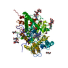
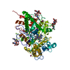
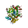
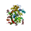
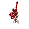
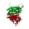
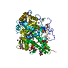
- PDB-1gne: THE THREE-DIMENSIONAL STRUCTURE OF GLUTATHIONE S-TRANSFERASE OF S... -
+ Open data

- Basic information
| Entry | Database: PDB / ID: 1gne | ||||||
|---|---|---|---|---|---|---|---|
| Title | THE THREE-DIMENSIONAL STRUCTURE OF GLUTATHIONE S-TRANSFERASE OF SCHISTOSOMA JAPONICUM FUSED WITH A CONSERVED NEUTRALIZING EPITOPE ON GP41 OF HUMAN IMMUNODEFICIENCY VIRUS TYPE 1 | ||||||
 Components Components | GLUTATHIONE S-TRANSFERASE | ||||||
 Keywords Keywords | TRANSFERASE / GLUTATHIONE TRANSFERASE | ||||||
| Function / homology |  Function and homology information Function and homology information | ||||||
| Biological species | synthetic construct (others) | ||||||
| Method | X-RAY DIFFRACTION / Resolution: 2.5 Å | ||||||
 Authors Authors | Lim, K. / Ho, J.X. / Keeling, K. / Gilliland, G.L. / Ji, X. / Ruker, F. / Carter, D.C. | ||||||
 Citation Citation | Journal: Protein Sci. / Year: 1994 Title: Three-dimensional structure of Schistosoma japonicum glutathione S-transferase fused with a six-amino acid conserved neutralizing epitope of gp41 from HIV. Authors: Lim, K. / Ho, J.X. / Keeling, K. / Gilliland, G.L. / Ji, X. / Ruker, F. / Carter, D.C. #1: Journal: PROTEIN PEPT.LETT. / Year: 1994Title: Fusion Proteins as Alternate Crystallization Paths to Difficult Structure Problems Authors: Carter, D.C. / Ruker, F. / Ho, J.X. / Lim, K. / Keeling, K. / Gilliland, G.L. / Ji, X. #2: Journal: J.Mol.Biol. / Year: 1994Title: Molecular Structure at 1.8 Angstroms of Mouse Liver Class Pi Glutathione S-Transferase Complexed with S-(P-Nitrobenzyl)Glutathione and Other Inhibitors Authors: Garcia-Saez, I. / Parraga, A. / Phillips, M.F. / Mantle, T.J. / Coll, M. #4: Journal: J.Virol. / Year: 1993Title: A Conserved Neutralizing Epitope on Gp41 of Human Immunodeficiency Virus Type 1 Authors: Muster, T. / Steindl, F. / Purtscher, M. / Trkola, A. / Klima, A. / Himmler, G. / Ruker, F. / Katinger, H. #5: Journal: J.Mol.Biol. / Year: 1993Title: Structure Determination and Refinement of Human Alpha Class Glutathione Transferase A1-1, and a Comparison with the Mu and Pi Class Enzymes Authors: Sinning, I. / Kleywegt, G.J. / Cowan, S.W. / Reinemer, P. / Dirr, H.W. / Huber, R. / Gilliland, G.L. / Armstrong, R.N. / Ji, X. / Board, P.G. / Olin, B. / Mannervik, B. / Jones, T.A. #6: Journal: Biochemistry / Year: 1992Title: The Three-Dimensional Structure of a Glutathione S-Transferase from the Mu Gene Class. Structural Analysis of the Binary Complex of Isoenzyme 3-3 and Glutathione at 2.2 Angstroms Resolution Authors: Ji, X. / Zhang, P. / Armstrong, R.N. / Gilliland, G.L. #7: Journal: J.Mol.Biol. / Year: 1992Title: Three-Dimensional Structure of Class P Glutathione S-Transferase from Human Placenta in Complex with S-Hexylglutathione at 2.8 Angstroms Resolution Authors: Reinemer, P. / Dirr, H.W. / Ladenstein, R. / Huber, R. / Lobello, M. / Federici, G. / Parker, M.W. #8: Journal: Embo J. / Year: 1991Title: The Three-Dimensional Structure of Class P Glutathione S-Transferase in Complex with Glutathione Sulfonate at 2.3 Angstroms Resolution Authors: Reinemer, P. / Dirr, H.W. / Ladenstein, R. / Schiffer, J. / Gallay, O. / Huber, R. #9: Journal: Gene / Year: 1988Title: Single-Step Purification of Polypeptides Expressed in Escherichia Coli as Fusions with Glutathione S-Transferase Authors: Smith, D.B. / Johnson, K.S. #10: Journal: Proc.Natl.Acad.Sci.USA / Year: 1987Title: Correction Authors: Smith, D.B. / Davern, K.M. / Board, P.G. / Tiu, W.U. / Garcia, E.G. / Mitchell, G.F. #11: Journal: Proc.Natl.Acad.Sci.USA / Year: 1986Title: Mr 26,000 Antigen of Schistosoma Japonicum Recognized by Resistant Wehi 129(Slash)J Mice is a Parasite Glutathione S-Transferase Authors: Smith, D.B. / Davern, K.M. / Board, P.G. / Tiu, W.U. / Garcia, E.G. / Mitchell, G.F. | ||||||
| History |
|
- Structure visualization
| Structure viewer | Molecule: MolmilJmol/JSmol |
|---|
- Downloads & links
-Download
| PDBx/mmCIF format | 1gne.cif.gz | 64.7 KB | Display | PDBx/mmCIF format |
|---|---|---|---|---|
| PDB format | pdb1gne.ent.gz | 46.4 KB | Display | PDB format |
| PDBx/mmJSON format | 1gne.json.gz | Tree view | PDBx/mmJSON format | |
| Others |  Other downloads Other downloads |
-Validation report
| Arichive directory | https://data.pdbj.org/pub/pdb/validation_reports/gn/1gneftp://data.pdbj.org/pub/pdb/validation_reports/gn/1gne | HTTPS FTP |
|---|
-Related structure data
| Similar structure data |
|---|


-Links
 PDBj
PDBj

- Assembly
| Deposited unit | 
| ||||||||
|---|---|---|---|---|---|---|---|---|---|
| 1 | 
| ||||||||
| Unit cell |
| ||||||||
| Atom site foot note | 1: CIS PROLINE - PRO 55 / 2: CIS PROLINE - PRO 201 3: HIS 214 - PRO 215 OMEGA = 246.61 PEPTIDE BOND DEVIATES SIGNIFICANTLY FROM TRANS CONFORMATION |
-Components
| #1: Protein | Mass: 27091.445 Da / Num. of mol.: 1 Source method: isolated from a genetically manipulated source Source: (gene. exp.) synthetic construct (others) / Gene: GP41 / Organ: LIVER / Gene (production host): GP41 / References: UniProt: P08515, glutathione transferase |
|---|---|
| #2: Chemical | ChemComp-GSH / Glutathione  Mass: 307.323 Da / Num. of mol.: 1 / Source method: obtained synthetically / Formula: C10H17N3O6S Mass: 307.323 Da / Num. of mol.: 1 / Source method: obtained synthetically / Formula: C10H17N3O6S |
| #3: Water | ChemComp-HOH / Water Mass: 18.015 Da / Num. of mol.: 126 / Source method: isolated from a natural source / Formula: H2O Mass: 18.015 Da / Num. of mol.: 126 / Source method: isolated from a natural source / Formula: H2O |
-Experimental details
-Experiment
| Experiment | Method: X-RAY DIFFRACTION |
|---|
- Sample preparation
| Crystal | Density Matthews: 2.41 Å3/Da / Density % sol: 48.88 % | |||||||||||||||||||||||||
|---|---|---|---|---|---|---|---|---|---|---|---|---|---|---|---|---|---|---|---|---|---|---|---|---|---|---|
| Crystal grow | *PLUS Temperature: 22 ℃ / pH: 6.5 / Method: vapor diffusion, hanging drop | |||||||||||||||||||||||||
| Components of the solutions | *PLUS
|
-Data collection
| Radiation | Scattering type: x-ray |
|---|---|
| Radiation wavelength | Relative weight: 1 |
| Reflection | *PLUS Highest resolution: 2.5 Å / Num. obs: 8485 / % possible obs: 86.9 % / Observed criterion σ(I): 1 / Rmerge(I) obs: 0.117 |
- Processing
| Software |
| ||||||||||||||||||||||||||||||||||||||||||||||||||||||||||||
|---|---|---|---|---|---|---|---|---|---|---|---|---|---|---|---|---|---|---|---|---|---|---|---|---|---|---|---|---|---|---|---|---|---|---|---|---|---|---|---|---|---|---|---|---|---|---|---|---|---|---|---|---|---|---|---|---|---|---|---|---|---|
| Refinement | Resolution: 2.5→6 Å / σ(F): 2 /
| ||||||||||||||||||||||||||||||||||||||||||||||||||||||||||||
| Refinement step | Cycle: LAST / Resolution: 2.5→6 Å
| ||||||||||||||||||||||||||||||||||||||||||||||||||||||||||||
| Refine LS restraints |
| ||||||||||||||||||||||||||||||||||||||||||||||||||||||||||||
| Software | *PLUS Name: X-PLOR / Classification: refinement | ||||||||||||||||||||||||||||||||||||||||||||||||||||||||||||
| Refinement | *PLUS Rfactor obs: 0.219 | ||||||||||||||||||||||||||||||||||||||||||||||||||||||||||||
| Solvent computation | *PLUS | ||||||||||||||||||||||||||||||||||||||||||||||||||||||||||||
| Displacement parameters | *PLUS | ||||||||||||||||||||||||||||||||||||||||||||||||||||||||||||
| Refine LS restraints | *PLUS
|
