 Movie
Movie Controller


+ Open data

- Basic information
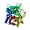
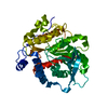
| Entry | Database: PDB / ID: 1g2p | ||||||
|---|---|---|---|---|---|---|---|
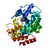
| Title | CRYSTAL STRUCTURE OF ADENINE PHOSPHORIBOSYLTRANSFERASE | ||||||
 Components Components | ADENINE PHOSPHORIBOSYLTRANSFERASE 1 | ||||||
 Keywords Keywords | TRANSFERASE / dimer / catalytic loop | ||||||
| Function / homology |  Function and homology information Function and homology informationPurine salvage / adenine binding / adenine salvage / adenine phosphoribosyltransferase activity / adenine phosphoribosyltransferase / AMP salvage / purine ribonucleoside salvage / AMP binding / Neutrophil degranulation / metal ion binding ...Purine salvage / adenine binding / adenine salvage / adenine phosphoribosyltransferase activity / adenine phosphoribosyltransferase / AMP salvage / purine ribonucleoside salvage / AMP binding / Neutrophil degranulation / metal ion binding / nucleus / cytoplasmSimilarity search - Function | ||||||
| Biological species |  Saccharomyces cerevisiae (brewer's yeast) Saccharomyces cerevisiae (brewer's yeast) | ||||||
| Method | X-RAY DIFFRACTION / SYNCHROTRON / MOLECULAR REPLACEMENT / Resolution: 1.75 Å | ||||||
 Authors Authors | Shi, W. / Tanaka, K.S.E. / Almo, S.C. / Schramm, V.L. | ||||||
 Citation Citation | Journal: Biochemistry / Year: 2001 Title: Structural analysis of adenine phosphoribosyltransferase from Saccharomyces cerevisiae. Authors: Shi, W. / Tanaka, K.S. / Crother, T.R. / Taylor, M.W. / Almo, S.C. / Schramm, V.L. | ||||||
| History |
|
- Structure visualization
| Structure viewer | Molecule: MolmilJmol/JSmol |
|---|
- Downloads & links
-Download
| PDBx/mmCIF format | 1g2p.cif.gz | 47.9 KB | Display | PDBx/mmCIF format |
|---|---|---|---|---|
| PDB format | pdb1g2p.ent.gz | 33.7 KB | Display | PDB format |
| PDBx/mmJSON format | 1g2p.json.gz | Tree view | PDBx/mmJSON format | |
| Others |  Other downloads Other downloads |
-Validation report
| Arichive directory | https://data.pdbj.org/pub/pdb/validation_reports/g2/1g2pftp://data.pdbj.org/pub/pdb/validation_reports/g2/1g2p | HTTPS FTP |
|---|
-Related structure data
| Related structure data | 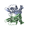 1g2qC 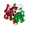 1qb8S S: Starting model for refinement C: citing same article ( |
|---|---|
| Similar structure data |


-Links
 PDBj
PDBj


- Assembly
| Deposited unit | 
| ||||||||
|---|---|---|---|---|---|---|---|---|---|
| 1 | 
| ||||||||
| Unit cell |
| ||||||||
| Details | The second part of the biological assemble is generated by the two fold screw axis, -x, -y+1/2, z |
-Components
| #1: Protein | Mass: 20616.814 Da / Num. of mol.: 1 Source method: isolated from a genetically manipulated source Source: (gene. exp.) Saccharomyces cerevisiae (brewer's yeast)Plasmid: PQE / Production host:  Escherichia coli (E. coli) Escherichia coli (E. coli)References: UniProt: P49435, adenine phosphoribosyltransferase | ||
|---|---|---|---|
| #2: Chemical | Sulfate  Mass: 96.063 Da / Num. of mol.: 2 / Source method: obtained synthetically / Formula: SO4 Mass: 96.063 Da / Num. of mol.: 2 / Source method: obtained synthetically / Formula: SO4#3: Water | ChemComp-HOH / | Water Mass: 18.015 Da / Num. of mol.: 108 / Source method: isolated from a natural source / Formula: H2O Mass: 18.015 Da / Num. of mol.: 108 / Source method: isolated from a natural source / Formula: H2O |
-Experimental details
-Experiment
| Experiment | Method: X-RAY DIFFRACTION / Number of used crystals: 1 |
|---|
- Sample preparation
| Crystal | Density Matthews: 3.19 Å3/Da / Density % sol: 61 % | ||||||||||||||||||||||||
|---|---|---|---|---|---|---|---|---|---|---|---|---|---|---|---|---|---|---|---|---|---|---|---|---|---|
| Crystal grow | Temperature: 298 K / Method: vapor diffusion, hanging drop / pH: 7.5 Details: lithium sulfate, Hepes, pH 7.5, VAPOR DIFFUSION, HANGING DROP, temperature 298K | ||||||||||||||||||||||||
| Crystal grow | *PLUS Temperature: 18 ℃ / Method: vapor diffusion | ||||||||||||||||||||||||
| Components of the solutions | *PLUS
|
-Data collection
| Diffraction | Mean temperature: 100 K |
|---|---|
| Diffraction source | Source: SYNCHROTRON / Site: NSLS  / Beamline: X9B / Wavelength: 0.98 Å / Beamline: X9B / Wavelength: 0.98 Å |
| Detector | Type: ADSC QUANTUM 4 / Detector: CCD / Date: Jul 11, 1999 |
| Radiation | Protocol: SINGLE WAVELENGTH / Monochromatic (M) / Laue (L): M / Scattering type: x-ray |
| Radiation wavelength | Wavelength: 0.98 Å / Relative weight: 1 |
| Reflection | Resolution: 1.75→20 Å / Num. all: 26348 / Num. obs: 26348 / % possible obs: 95.5 % / Observed criterion σ(F): 0 / Observed criterion σ(I): 0 / Redundancy: 3.9 % / Biso Wilson estimate: 15.9 Å2 / Rsym value: 0.041 / Net I/σ(I): 29.6 |
| Reflection shell | Resolution: 1.75→1.78 Å / Redundancy: 1.9 % / Mean I/σ(I) obs: 3.8 / Num. unique all: 1134 / Rsym value: 0.24 / % possible all: 82.8 |
| Reflection | *PLUS Num. measured all: 103449 / Rmerge(I) obs: 0.041 |
| Reflection shell | *PLUS % possible obs: 82.8 % / Rmerge(I) obs: 0.24 / Mean I/σ(I) obs: 2.6 |
- Processing
| Software |
| ||||||||||||||||||||||||||||||||||||||||
|---|---|---|---|---|---|---|---|---|---|---|---|---|---|---|---|---|---|---|---|---|---|---|---|---|---|---|---|---|---|---|---|---|---|---|---|---|---|---|---|---|---|
| Refinement | Method to determine structure: MOLECULAR REPLACEMENT Starting model: pdb entry 1QB8 Resolution: 1.75→20 Å / Rfactor Rfree error: 0.005 / Isotropic thermal model: restrained / Cross valid method: THROUGHOUT / σ(F): 2 / σ(I): 1.4 / Stereochemistry target values: Engh & Huber
| ||||||||||||||||||||||||||||||||||||||||
| Solvent computation | Solvent model: Flat model / Bsol: 49.4464 Å2 / ksol: 0.370397 e/Å3 | ||||||||||||||||||||||||||||||||||||||||
| Displacement parameters | Biso mean: 28.6 Å2
| ||||||||||||||||||||||||||||||||||||||||
| Refine analyze |
| ||||||||||||||||||||||||||||||||||||||||
| Refinement step | Cycle: LAST / Resolution: 1.75→20 Å
| ||||||||||||||||||||||||||||||||||||||||
| Refine LS restraints |
| ||||||||||||||||||||||||||||||||||||||||
| LS refinement shell | Resolution: 1.75→1.86 Å / Rfactor Rfree error: 0.018 / Total num. of bins used: 6
| ||||||||||||||||||||||||||||||||||||||||
| Software | *PLUS Name: CNS / Version: 0.9 / Classification: refinement | ||||||||||||||||||||||||||||||||||||||||
| Refinement | *PLUS Lowest resolution: 20 Å / σ(F): 2 / % reflection Rfree: 9.8 % / Rfactor all: 0.21 / Rfactor obs: 0.208 / Rfactor Rfree: 0.23 | ||||||||||||||||||||||||||||||||||||||||
| Solvent computation | *PLUS | ||||||||||||||||||||||||||||||||||||||||
| Displacement parameters | *PLUS Biso mean: 28.6 Å2 | ||||||||||||||||||||||||||||||||||||||||
| Refine LS restraints | *PLUS
| ||||||||||||||||||||||||||||||||||||||||
| LS refinement shell | *PLUS Rfactor Rfree: 0.329 / % reflection Rfree: 9.8 % / Rfactor Rwork: 0.3 |
